Author: Aleksandra Zurowska, EMJ, London, UK
Citation: EMJ. 2026;11[1]:10-13. https://doi.org/10.33590/emj/3Y43909F
![]()
AS DATASETS expand and computational tools mature, hepatopancreatobiliary medicine is entering a new phase in which AI is no longer experimental, but increasingly embedded in research and clinical strategy. During the session ‘What’s new in AI and big data in HPB in 2025?’ at the United European Gastroenterology (UEG) Week 2025 in Berlin, Germany, speakers explored how predictive modelling, electronic health record integration, and multi-omics data are redefining risk prediction, surgical planning, and long-term disease surveillance.
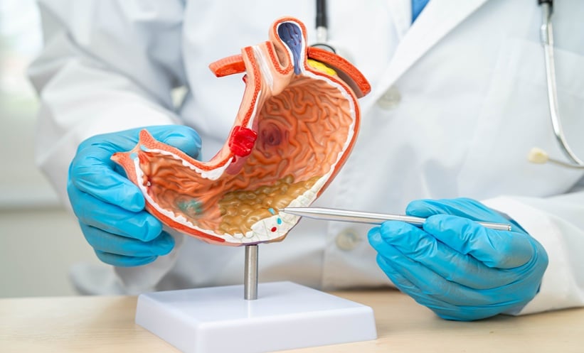
AI IN PANCREATIC SURGERY
AI is rapidly reshaping the landscape of surgical oncology. However, its true potential lies not in isolated tools, but rather in the integration of multiple data layers. The talk delivered by Andrew Gumbs, Hôpital Antoine-Béclère, Clamart, France, outlined how AI-driven multi-omics approaches, including radiomics, pathomics, genomics, and surgical video analysis, could redefine personalised cancer care, particularly in pancreatic adenocarcinoma.
Building his argument around the Artificial intelligence, Radiomics, Genomics, Oncopathomics and Surgomics (AiRGOS) project, a pan-European initiative aimed at combining imaging, pathology, genomic, and intraoperative visual data in surgery across multiple centres, he explained how the ambition is to move beyond static tumour boards towards AI-supported decision-making, including real-time guidance in the operating room. However, while the computational tools already exist, Gumbs emphasised that legal and regulatory barriers within the EU remain the greatest obstacle to progress, often preventing effective data sharing between institutions.
Radiomics was highlighted as one of the most mature AI applications in oncology. By analysing tumours voxel-by-voxel in 3D, radiomics enables extraction of shape, intensity, and texture features far beyond the human visual capacity. When paired with deep learning, these techniques have demonstrated the ability to stratify tumours by biological behaviour, including early versus late recurrence, even from a single imaging phase. While such models often function as “black boxes,” Gumbs argued that this should not preclude their clinical use, provided AI remains an advisor rather than an autonomous decision-maker.
Pathomics, an emerging approach that applies AI and computer vision to digitised histology slides to extract quantitative data on tumour morphology, has increasingly gained attention and may currently represent the most powerful predictive omics in pancreatic cancer. Emerging multicentre data suggest that deep learning applied to pathology images can capture tumour biology with remarkable precision, potentially rivalling or even surpassing genomic approaches in certain contexts.1 Although whole-genome sequencing has become significantly more affordable, its widespread adoption is still constrained by access and funding rather than cost alone.1
Gumbs also addressed ethical considerations around data ownership and consent. Blockchain-based solutions were proposed as a way to give patients transparency and agency over how their data are used, offering a potential framework for fair and traceable data sharing in the future.
In closing, Gumbs stressed that the future of oncology lies in multi-omics integration, not single modality analysis. While Europe risks falling behind due to its regulations, scientific societies and international collaborations are well placed to drive progress. Ultimately, AI-enabled tumour boards could help clinicians move away from trial-and-error treatment strategies towards truly biologically informed, personalised cancer care.
BIG DATA AND AI IN PERSONALISED THERAPY IN METABOLIC LIVER DISEASES
Rising Star awardee Carolin Schneider, RWTH Aachen University Hospital, Germany, continued the session with a talk on harnessing big data and AI to enable personalised therapy in metabolic liver diseases.
Schneider began by framing the scale of the problem: obesity and metabolic liver disease now affect approximately 30% of the global population.2,3 The stepwise progression from steatosis to steatohepatitis, cirrhosis, and ultimately hepatocellular carcinoma presents clear windows for early intervention. However, she emphasised that the pathogenesis of liver disease is multidimensional, shaped by lifestyle, genetics, sex, medication exposure, and broader societal factors.
To address this complexity, Schneider described an ambitious, data-driven research programme built around harmonised international cohorts (Schneider, unpublished data). Collectively, these datasets include more than three million individuals with a mean follow-up of approximately 14 years. Beyond standard clinical information, the cohorts contain detailed demographic and lifestyle data, electronic health records, routine serum biomarkers, whole-genome data, and, in some cases, more than 250 metabolomic parameters. Liver disease serves as the starting point, but the infrastructure is designed to expand into wider gastrointestinal and metabolic conditions.
One key line of investigation focused on lifestyle behaviour. In a cohort of over 100,000 individuals who wore fitness trackers for 1 year, daily step counts were analysed in relation to future metabolic liver disease risk.4 Schneider reported that approximately 7,500 steps per day were associated with a meaningful reduction in risk, while 12,000 steps were linked to roughly halving the risk over 3.5 years. The findings suggest that protective behavioural targets may be more attainable than often assumed.4
Nutrition was examined in more than 210,000 individuals who completed repeated 24-hour dietary questionnaires.5 Higher vitamin E intake was associated with lower liver disease risk, supporting previous clinical observations. When machine learning techniques were applied, using a random forest model trained on 64 nutrients, manganese emerged as a leading associated factor. Higher manganese intake correlated with lower liver disease incidence, even after adjustment for confounders. However, Schneider cautioned that manganese-rich foods, such as nuts and whole grains, may simply reflect an overall healthier dietary pattern, highlighting the hypothesis-generating nature of such analyses.5
Turning to hepatocellular carcinoma, Schneider presented work leveraging UK Biobank data to develop predictive models of increasing complexity. The team described a series of decision tree-based approaches, beginning with anthropometric and lifestyle variables and progressively incorporating electronic health records, laboratory markers, genetic variants, and metabolomic profiles. While the most comprehensive model delivered the highest predictive performance, Schneider highlighted that routine serum biomarkers alone demonstrated substantial predictive value, underscoring the clinical promise of readily available data.
A key focus of the presentation was model bias. Analysis revealed marked sex-based disparities: the algorithm identified 73% of male hepatocellular carcinoma cases, but only 31% of female cases. Schneider attributed this imbalance to skewed training data and stressed the necessity of subgroup validation and bias-aware model development to avoid perpetuating existing inequities in cancer detection.
Schneider concluded that, while AI offers powerful tools for hepatology, validation, transparency, and bias awareness remain essential. Precision prevention, she suggested, will depend not only on technological innovation, but on careful and equitable implementation. Robust associations validated across multiple cohorts may justify targeted prospective trials with higher probabilities of success. Synthetic trial methodologies and cross-cohort transfer learning are emerging strategies, but conventional randomised evidence remains essential.
Future directions include integrating tabular data with imaging modalities, such as ultrasound, to enhance hepatocellular carcinoma risk stratification. Schneider also highlighted ongoing efforts in cross-cohort transfer learning, enabling knowledge derived from large datasets to inform smaller, deeply characterised cohorts, and vice versa.
Ultimately, her message was both ambitious and grounded: precision hepatology will require scale, collaboration, and vigilance. In Berlin, big data was not presented as a futuristic abstraction, but as an evolving infrastructure already reshaping how metabolic liver disease is understood and, potentially, prevented.
AI IMAGING FOR PANCREATIC DISEASES
Closing off the session at UEG Week 2025, Adrian Saftoiu, University of Medicine and Pharmacy of Craiova, Romania, delivered the final talk on the role of AI as an imaging tool in pancreatic diseases, moving from risk prediction models to real-time endoscopic ultrasound (EUS) guidance and emerging applications in pathology and robotics.
Saftoiu began by explaining the central role of EUS in pancreatic cancer diagnosis. Beyond imaging, EUS enables fine-needle aspiration biopsy for tissue confirmation and supports screening and prognostic assessment. However, with newer techniques such as Doppler flow imaging, contrast harmonic imaging, and elastography, the learning curve has grown increasingly steep. AI, he suggested, offers a way to shorten that curve.
He highlighted large-scale screening efforts such as the Pancreatic Duct Adenocarcinoma Risk Model (PRISM), built using over 1.5 million controls and 35,000 pancreatic ductal adenocarcinoma cases. By integrating clinical data, biomarkers, and imaging features, the model identified high-risk individuals beyond those captured by current guidelines, potentially expanding early detection strategies.6
In CT imaging, Saftoiu referenced work demonstrating that non-contrast-enhanced scans can detect pancreatic malignancies with performance comparable to radiologists using contrast-enhanced studies.7 Such findings suggest that AI may extract diagnostic value even from routinely acquired imaging.
Within EUS, earlier artificial neural network models modestly improved differentiation between chronic pancreatitis and pancreatic cancer. However, the field accelerated with convolutional neural networks. Saftoiu described a real-time EUS segmentation system trained on approximately 200 patients and validated on 300 more, capable of identifying pancreatic tissue, cysts, solid tumours, ducts, and even small stones without perceptible lag.8 Its accuracy matched expert operators, offering particular value for less experienced clinicians by guiding biopsy placement and ensuring complete examination.8
Digital pathology represents another advancing frontier. AI models applied to fine-needle aspiration biopsy slides showed near-perfect overlap with expert pathologist annotations in identifying adenocarcinoma regions. Integration with real-time imaging technologies could further streamline diagnosis.
Saftoiu also addressed large language models in clinical interpretation. While tools, such as ChatGPT (OpenAI, San Francisco, California, USA), can describe imaging features and suggest diagnoses, he cautioned that these probabilistic systems remain prone to error and require careful validation. In one clinical example, AI-supported interpretation was helpful but not definitive, reinforcing the need for clinical judgement.
Looking ahead, radiomics may allow AI to extract greyscale information beyond human visual perception, potentially enhancing differential diagnosis. However, questions of standardisation and generalisability remain. Saftoiu advocated for federated learning approaches and cloud-based platforms to enable multicentre collaboration and broader access.
He concluded that AI in pancreatic imaging is already embedded within current systems and will continue expanding into diagnostic support, therapy monitoring, and potentially robotic integration. The challenge now lies not in technological capability, but in rigorous validation and responsible implementation.