Hepatic stellate cells are responsible for liver tissue’s ability to store vitamin A and 80% of whole-body retinol as cytosolic lipid droplets of retinyl palmitate.1 They are distributed in the space between the parenchymal and sinusoidal cells.2 Hepatic stellate cells are physiologically quiescent and are characterised by epithelial morphology.
Liver insult, damage, and/or epigenetic modifications cause the transdifferentiation of stellate cells into myofibroblasts.3 Once the stellate cells transactivate into mesenchymal cells, they can infiltrate the liver parenchyma and are responsible for the collagen secretion, which can lead to liver fibrosis, cirrhosis, and tumourigenesis.3,4
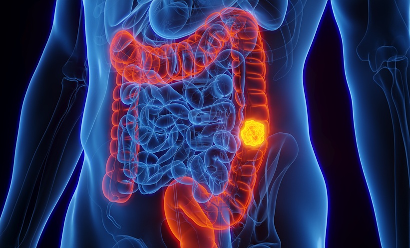
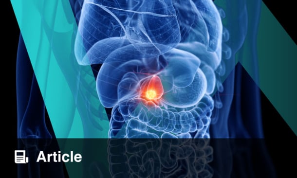
Pancreatic stellate cells (PSC) constitute the pancreas stroma. The cells carry out various functions, including the formation of the extracellular matrix, stimulation of amylase secretion, phagocytosis, and contributing to immunity.5 Quiescent PSC are characterised by vitamin A droplets; once active, they assume a myofibroblast-like morphology and are responsible for the fibrogenesis observed in pancreatitis and pancreatic ductal adenocarcinoma.6 Furthermore, it has been shown that PSC support tumour metabolism by inducing autophagic alanine secretion.7 Until now, the role exerted by PSC has been unclear, and it is not known if they exert a tumour-suppressing or a tumour-sustaining effect.
Our study highlights a possible intercommunication existing between the stellate cells, other pancreatic and liver cells, and the microbiota. In vitro studies show that LX28 and HPSC 2.29 immortalised stellate cells can be easily treated with low levels of tumour growth factor beta (TGF-β1), leading to their transdifferentiation into myofibroblasts. TGF-β1 was able to stimulate the stellate cells, promote their activation, and their change from a quiescent state to an active one, which characterises these cells in their liver and pancreatic tissue environment. The responsiveness to TGF-β1 highlights the fact that the stellate cells can be activated by signalling coming from both neighbouring and more distant cells. In particular, the presence of tumour cells in the surrounding parenchyma could be also responsible for stellate cell activation through the secretion of several growth stimulating factors, including TGF-β1.10
Currently, apart from their simple vitamin and fat storing role, the function of stellate cells is not yet fully understood; they could play a strategic role during tumourigenesis and promote intra and intercellular signalling, actively participating in the tumour environment.
Interestingly, TGF-β1 signalling was responsible in the proposed model for inducing the expression of toll-like receptor (TLR)5. This member of the TLR family is known for being easily activated by the bacterial protein flagellin. It was observed that knocking down the mRNA and protein level of TLR5 completely disabled TGF-β1 efficacy, leaving the liver and PSC in a quiescent state. The expression of TLR5 was essential for the stellate cells to acquire their active status and the efficacy of TGF-β1 was dispensable in cells knocked down for TLR5.
The importance of TLR5 in stellate cell activation offers new perspectives for understanding the mechanisms underlying stellate cells’ involvement in the tissue environment: in particular, the sensitivity of stellate cells to stimuli coming from micro-organisms populating the host body and the ability of micro-organisms to activate molecular and morphological changes in cells that play a specific role in the structure and functioning of the tissue or organ. Understanding the mechanisms of organ fibrogenesis in correlation with tumourigenesis and the influence of microbiota will open new directions for future treatment of diseases affecting the upper-midgut area of the human body.