BACKGROUND
Transcatheter mitral valve replacement (TMVR) using the HighLife TMVR system has demonstrated feasibility, an acceptable safety profile, and excellent reduction of mitral regurgitation, as well as functional improvement in symptomatic patients.1 Beyond correction of mitral regurgitation, the two-component HighLife technique potentially exerts annuloplasty by an undersizing docking mechanism, as opposed to several other approaches of anchoring which rely on outward radial force, which can lead to annular oversizing.2,3,4,5 The aim of this investigation was to assess annular remodelling after HighLife implantation using both echocardiographic studies as well as CT imaging.6
METHODS
Pre- and post-procedural transthoracic and procedural transoesophageal echocardiographic studies of 25 patients treated at University Heart Center Ulm (Germany) within the HighLife (HL2018-01-TS) and HighFLO (HL201-01) clinical trials were assessed by the site. The largest annular diameters were measured in three planes during diastole, both before and after valve implantation. Postprocedural CT was available in five patients.
RESULTS
Before valve implantation, measurements of anterior-posterior (A-P), anteromedial-posterolateral (AM-PL), and septal-lateral (S-L) diameters were comparable between transthoracic echocardiography (TTE) and transoesophageal echocardiography (TEE). A-P diameters were 40.2±5.4 mm and 39.6±4.9 mm, AM-PL diameters were 41.4±2.9 mm and 42.2±4.4 mm and S-L diameters were 38.9±3.1 mm and 39.5±3.2 mm in TTE and TEE, respectively. After valve implantation, TTE measurements were slightly larger: 37.5±4.9 mm (A-P), 37.0±2. 3mm (AM-PL), and 34.9±3.9 mm (S-L), compared to TEE: 35.5±4.5 mm (A-P), 38.0±3.4 mm (AM-PL), and 33.6±4.2 mm (S-L). However, both modalities showed significant reductions across all diameters, with pronounced effects on S-L dimensions (TTE: –6.1±9.4% A-P, –12.2±3.4% AM-PL, and –12.2±21.9% S-L, p=0.01 respectively; TEE: –9.9±9.9% A-P, –7.9±5.2% AM-PL, and –15.0±8.0% S-L, p<0.01 respectively; Figure 1). Assessment of available CT scans confirmed these echocardiographic findings.
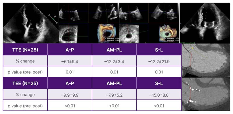
Figure 1: Changes of mitral annular dimension pre- and post-intervention measured in transoesophageal and transthoracic echocardiography.
A-P: anterior-posterior; AM-PL: anteromedial-posterolateral; S-L: septal-lateral; TEE: transoesophageal echocardiography; TTE: transthoracic echocardiography.
CONCLUSION
Annular measurements of a relatively small group of patients treated at one German site within the HighLife and HighFLO clinical trials showed significant annuloplasty independent of eliminating mitral regurgitation. A larger cohort should be assessed including CT assessment to confirm these findings and correlate annuloplasty with ventricular remodeling and outcome.