ADVANCED fetal imaging performed at 34-38 weeks’ gestation helped clinicians identify babies with dextro-transposition of the great arteries (d-TGA) who were most likely to experience severe hypoxia after birth and require urgent cardiac intervention, a prospective cohort study found.
What d-TGA Means for Newborns
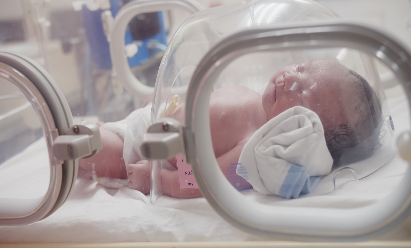
d-TGA is a congenital heart defect in which the aorta and the pulmonary artery are reversed. This abnormal arrangement limits the mixing of oxygen-rich and oxygen-poor blood in the heart, placing newborns at risk of critical oxygen deprivation immediately after delivery. In severe cases, clinicians performed balloon atrial septostomy (BAS) within 3 hours of birth to improve blood mixing between the heart’s upper chambers.
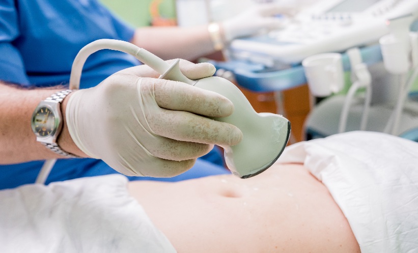
How Fetal Imaging Identified High-Risk Babies
The study included 34 pregnancies in which fetuses had d-TGA with either an intact ventricular septum or a small ventricular septal defect of 4 mm or less. All fetuses underwent fetal echocardiography, using two-dimensional imaging and Doppler to examine cardiac structures, including the atrial septum and foramen ovale.
Where feasible, cardiac magnetic resonance imaging (CMR) was performed concurrently to quantify fetal weight, brain volume, and vascular oxygen saturation. Both imaging methods assessed fetal blood flow in room air and during acute maternal hyperoxygenation (AMH), when the mother breathed supplemental oxygen to temporarily increase fetal oxygen levels.
Imaging Markers Predicted Urgent Intervention
Following delivery, 23 of 34 newborns underwent BAS, including 12 infants who required urgent intervention within three hours because preductal oxygen saturation remained below 70%.
Several imaging features were strongly associated with urgent BAS. On fetal echocardiography, abnormal atrial septal flap morphology, effective foramen ovale size ≤3.4 mm during AMH, and constricted or continuously reversed diastolic flow across the ductus arteriosus during AMH were the most significant predictors. On CMR, fetuses with an ascending aorta to main pulmonary artery oxygen saturation ratio >1 during AMH were significantly more likely to undergo BAS postnatally.
Implications For Prenatal Cardiac Assessment
The findings suggested that combining fetal echocardiography with CMR could improve prenatal risk assessment in pregnancies affected by d-TGA. By identifying high-risk babies before delivery, clinicians could better plan immediate postnatal care and ensure access to urgent cardiac interventions.
Although the study involved a small single-centre cohort, the results highlighted the value of late-gestation fetal imaging in anticipating neonatal hypoxia and guiding clinical management.
Reference
Jaeggi E et al. Role of predelivery fetal echocardiography, cardiac magnetic resonance imaging and acute maternal hyperoxygenation in predicting urgency of neonatal balloon atrial septostomy in fetal d-transposition of the great arteries. Ultrasound Obstet Gynecol. 2026; DOI:10.1002/uog.70200
Featured image: HarryKiiM Stock on Adobe Stock