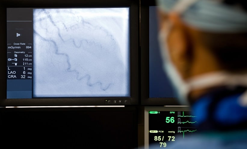
A RECENT study has demonstrated that digital variance angiography (DVA) significantly reduces radiation exposure while maintaining, and even improving, image quality during endovascular peripheral interventions (EPIs). With radiation dose reduction being a key objective in interventional radiology, this investigation offers a valuable alternative to conventional digital subtraction angiography (DSA). The findings are especially relevant for patients undergoing procedures on the lower extremities, where repeated imaging can pose cumulative radiation risks. Crucially, DVA reduced radiation dose by over 50% across all anatomical regions studied.
To assess the effectiveness of DVA in lowering radiation exposure, researchers evaluated its use during EPIs using version 6.0.4 of the Kinepict Medical Imaging Tool. Standard dose (EPI-ND) protocols delivering 1.20–0.81 µGy/frame were compared with low-dose (EPI-LD) protocols using DVA, which operated at 0.36 µGy/frame and in some cases 0.24 µGy/frame. A total of 432 interventions were included: 370 using standard dose protocols and 62 with DVA-based low dose protocols. The dose area product (DAP) was recorded to assess radiation exposure, while the contrast-to-noise ratio (CNR) was measured to evaluate image quality.
Statistical analysis revealed that DVA-LD protocols reduced median DAP significantly across all targeted regions: 62.0% in the pelvic region, 53.8% in the femoral and popliteal arteries, and 59.4% in cruro-pedal segments (p<0.005). Furthermore, DVA-LD achieved a significantly higher CNR than conventional low-dose DSA (p<0.001) and matched the image quality of normal-dose DSA (p>0.15). Relative improvements in CNR with DVA-LD compared to DSA-ND were notable, with enhancement ratios of 1.9 in the pelvic area and 2.4 in both femoral-popliteal and cruro-pedal regions.
This study supports the use of DVA as an effective imaging tool for reducing patient radiation exposure in lower extremity EPIs without compromising diagnostic quality. The findings suggest a valuable shift towards low-dose imaging protocols in clinical practice. However, the relatively small sample size for the low-dose group and the single-centre nature of the study may limit generalisability. Further research across multiple centres and patient populations would help validate these promising results and guide widespread adoption.
Reference
Schürmann T et al. Radiation exposure reduction in peripheral interventions using digital variance angiography versus conventional angiography. Nature. 2025;15(1):24693.


