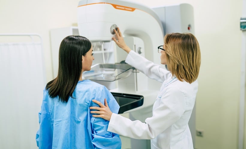
Breast cancer screening programmes rely heavily on mammography to identify potential cases, but the complexity of tissue structures and their variation across individuals can impact the accuracy of detection. A recent study explored the role of parenchymal phenotypes and their relationship with the future risk of breast cancer. The primary aim was to assess whether parenchymal phenotypes, derived from radiomic texture features from full-field digital mammography (FFDM), could predict future risk of breast cancer and the occurrence of masking (false-negative findings or interval cancers). Notably, the study also evaluated whether these phenotypes provide additional predictive value beyond traditional breast density classification. One key finding was that these phenotypes were significantly linked to a higher risk of breast cancer, as well as to false-negative findings and symptomatic interval cancers.
The study used a two-stage design, combining a retrospective cross-sectional study of 30,000 women, with a nested case-control study involving 1055 women with invasive breast cancer, matched to 2764 women without breast cancer. Radiomic features were extracted from FFDM images, with 390 features analysed and adjusted for age and practice differences. Hierarchical clustering and principal component analysis (PCA) were applied to classify the phenotypes, and these were then examined for associations with breast cancer risk, false-negative findings, and symptomatic interval cancers using conditional logistic regression. Discrimination for breast cancer was assessed through the area under the receiver operating characteristic curve (AUC).
The results revealed that six distinct clusters and six principal components (PCs) of parenchymal phenotypes were associated with a significantly higher risk of invasive breast cancer (p=0.01 and p<0.001, respectively). These PCs were also positively correlated with an increased likelihood of false-negative mammogram findings (p=0.004) and symptomatic interval cancers (p=0.006). When PCs were incorporated into risk models, there was a noticeable improvement in predicting false-negative findings (AUC 0.73 vs 0.66) and symptomatic interval cancers (AUC 0.77 vs 0.68), with both improvements being statistically significant (p=0.004 and p=0.006, respectively).
This study highlights the potential clinical value of incorporating parenchymal phenotypes derived from FFDM radiomics in breast cancer screening programmes. By providing additional risk information beyond traditional breast density classifications, these phenotypes could help improve early detection, particularly for women at higher risk of false-negative findings or interval cancers. However, the study is limited by its retrospective design and the generalisation of its findings across different populations. Further research, including prospective studies, would be beneficial to confirm these findings and assess their application in clinical practice.
Reference
Winham et al. Radiomic Parenchymal Phenotypes of Breast Texture from Mammography and Association with Risk of Breast Cancer. Radiology. 2025;DOI: 10.1148/radiol.240281.


