ACCURATE determination of HER-2 status in breast cancer is essential for guiding targeted therapy and improving clinical outcomes. While biopsy remains the gold standard, it is invasive and may not fully capture tumour heterogeneity. A recent study investigated whether combining radiomics features from different MRI modalities and tumour regions could non-invasively predict HER-2 expression. Notably, the combined model integrating peritumoral features from three MRI sequences showed the strongest predictive performance.
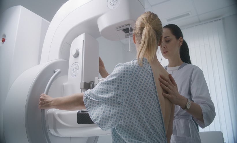
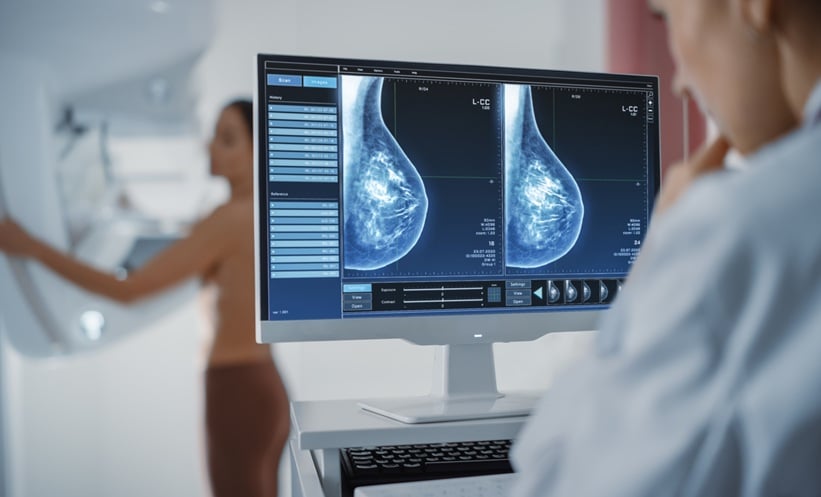
In the retrospective analysis of 266 women with pathologically confirmed breast cancer, researchers examined radiomics data from two medical centres. Patients from Centre 1 (n=199) were split into a training cohort (n=140) and internal validation cohort (n=59). An external test cohort was derived from Centre 2 (n=67). Tumours were segmented manually on T2-weighted, diffusion-weighted, and contrast-enhanced MRI. Radiomics features were extracted from both the tumour (intratumoral) and surrounding (peritumoral) tissue, with peritumoral volumes defined by a 3 mm expansion. Following feature selection, eight random forest models were trained to predict HER-2 status, and their performance evaluated through ROC analysis, calibration, and decision curve analysis.
The best-performing model combined peritumoral features from all three MRI sequences, DWI, T2WI, and DCE-MRI, and achieved high diagnostic accuracy. It yielded AUCs of 0.822; 95% CI: 0.755–0.889) in the training set, 0.823; 95% CI: 0.714–0.932) in the internal validation set, and 0.813; 95% CI: 0.712–0.914) in the external test set. This integrated model significantly outperformed models using single modalities or regions, demonstrating reliable generalisability across cohorts.
These findings support the clinical potential of radiomics-based MRI analysis in assessing HER-2 status non-invasively, which may assist in treatment planning and risk stratification. Importantly, the study highlights the added value of including peritumoral features, which may reflect tumour-host interactions and microenvironmental changes associated with HER-2 positivity. Limitations include its retrospective design, manual segmentation, and limited ethnic and geographic diversity, which may affect applicability in broader settings. Future prospective studies are warranted to validate these findings and assess their integration into routine diagnostic workflows.
Reference
Cao M et al. Prediction of HER-2 expression status in breast cancer based on multi-parameter MRI intratumoral and peritumoral radiomics. Magn Reson Imaging. 2025;DOI: 10.1016/j.mri.2025.110434.