MULTIPLEX MRI may support more detailed assessment of glioma grade and molecular subtype, according to new research exploring a single-scan quantitative imaging technique.
Glioma is a type of brain tumour that arises from glial cells in the central nervous system. It is broadly classified into lower-grade (grade 2/3) and higher-grade (grade 4) disease, with higher-grade tumours typically growing faster and associated with poorer survival. Accurate classification is essential for guiding treatment and estimating prognosis, particularly as modern approaches increasingly incorporate molecular markers alongside traditional histology.
What MULTIPLEX MRI Adds to Glioma Imaging
Standard MRI remains central to glioma evaluation but is largely based on visual interpretation. This qualitative approach can fall short when trying to reflect the molecular complexity now embedded in tumour classification. This is particularly important because distinct molecular subtypes can appear similar on conventional imaging.
MULTIPLEX MRI is an advanced quantitative imaging technique that captures multiple tissue measurements in a single scan. It generates numerical maps of key properties such as T1 and T2* relaxation times. T1 reflects how quickly the tissue returns to its normal state after being excited by the MRI signal, while T2* reflects how quickly the signal decays due to interactions within the tissue and local magnetic variations. These measurements give clinicians quantitative insight into tissue composition.
The technique also captures proton density, a measure of water content, and quantitative susceptibility mapping, which reflects magnetic characteristics of tissue. By combining these measurements, it provides a more detailed, data-driven picture of tumour biology compared with conventional imaging.
How the Study Assessed MULTIPLEX MRI
The study1 included 72 patients with diffuse glioma who underwent preoperative MRI. Researchers applied automated image processing to segment tumours into contrast-enhancing, oedematous, and necrotic regions.
From these areas, 48 quantitative parameters were derived using histogram-based analysis. Statistical analyses compared parameters between tumour grades and molecular subtypes.
Quantitative Differences Linked to Tumour Biology
The analysis identified measurable differences in imaging parameters across tumour groups. Lower-grade gliomas and tumours with certain molecular features, such as IDH mutation and MGMT methylation, showed higher average T2* values in necrotic or enhancing regions compared with higher-grade or less favourable subtypes.
Additional variations were observed in parameters reflecting distribution and variability. These included T1 skewness and variability in quantitative susceptibility mapping within necrotic tissue. Differences were also noted between tumours with and without 1p/19q co-deletion.
What These Findings Could Mean
These results suggest that MULTIPLEX MRI can capture quantitative differences associated with glioma grade and molecular characteristics. This approach may provide a more detailed, imaging-based view of tumour heterogeneity.
However, the findings are based on a relatively small cohort from a single centre and do not establish diagnostic performance or clinical benefit. These results do not replace biopsy or standard MRI, and further studies will be needed to determine how this approach might complement existing diagnostic pathways.
If validated, MULTIPLEX MRI could contribute to more integrated imaging assessments aligned with modern, molecularly informed glioma classification.
References
D Lin et al. The evaluation value of multiparametric imaging technology (MULTIPLEX) for grading and molecular subtyping of diffuse glioma patients. Clin Radiol. 2026;DOI:10.1016/j.crad.2026.107265.
S Azhar, LR Chong. Clinician’s guide to the basic principles of MRI. Postgrad Med J. 2022;99(1174):894-903.
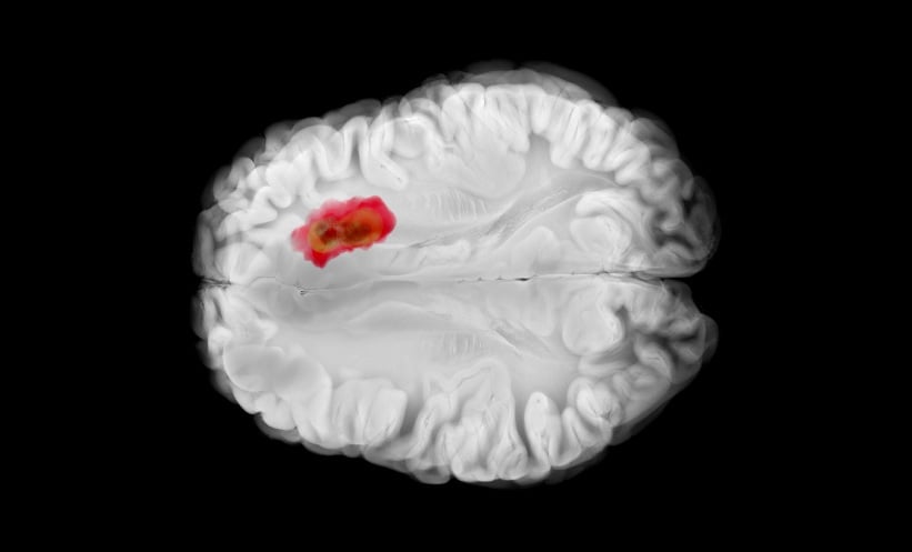
Featured image: Visual Voyager on Adobe stock