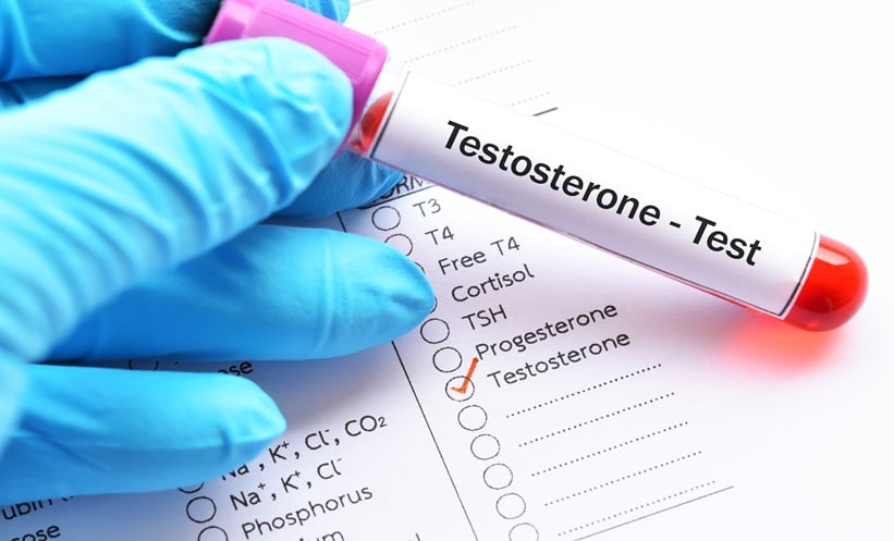
LOW SERUM testosterone has long been linked to more advanced prostate cancer at diagnosis and a higher risk of prostate cancer–specific mortality, a pattern plausibly explained by the androgen dependence of PSA expression: when testosterone is low, PSA often stays deceptively low, delaying referral for imaging and biopsy. Contemporary data from a prospectively followed, randomised PSA-screened cohort sharpen this concern.
After adjusting for age, clinical T-category and time-dependent use of salvage androgen deprivation therapy, individuals with low testosterone at presentation were more likely to harbour higher T-category disease and faced increased risks of prostate cancer–specific and all-cause mortality compared with peers with normal testosterone. Crucially, these signals were most evident among otherwise healthy participants, implying that under-detection rather than comorbidity burden is driving worse outcomes.
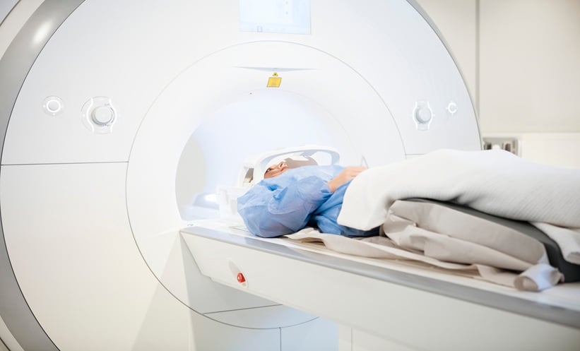
The diagnostic landscape has evolved as a standard 12-core transrectal ultrasound–guided biopsy can miss clinically significant tumours in about a third of cases, whereas combining multiparametric MRI with targeted and systematic sampling reduces that miss rate markedly. Yet screening guidance remains tethered to PSA thresholds, and routine digital rectal examination has receded from primary care. This poses a particular risk for men with low testosterone and transgender women receiving gender-affirming hormone therapy, in whom PSA is frequently very low; one cohort reported a median PSA of 0.02 ng/mL with over a third undetectable, meaning many cancers will not trigger investigation until late.
These observations justify testing an alternative pathway. A pragmatic trial could compare the current standard, annual PSA with MRI and biopsy only if PSA is elevated, against an investigational strategy of annual MRI plus PSA, proceeding to biopsy if either is abnormal. Stratification by transgender status and testosterone replacement would clarify whether biology or delayed detection predominates.
While MRI performance does not appear to depend on testosterone and inter-reader agreement is high, open questions remain, including whether PSMA PET or genomic markers could further refine early detection and which PSA kinetics, if any, warrant MRI in low-testosterone populations. Taken together, the available evidence suggests the hazard lies less in inherently aggressive biology and more in a PSA-led blind spot that MRI-driven screening might responsibly close.
Reference
Fattahi S et al. Low testosterone level and mortality risk in patients with prostate cancer: a post-randomization analysis. Cancer Med. 2025;14(15):e71124.