ULTRA-HIGH-FIELD 5 Tesla (T) MRI delivers superior image quality and improved diagnostic performance compared with standard 3 T MRI for prostate cancer (PCa), according to a prospective intra-individual study.
The study is among the first to directly compare 5 T and 3 T prostate MRI within the same patients, providing early clinical evidence for the feasibility and potential advantages of ultra-high-field imaging in prostate cancer assessment.
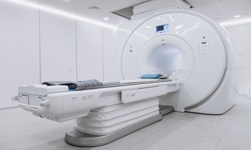
Comparing 5 T and 3 T MRI for Prostate Cancer Assessment
Researchers enrolled 67 consecutive patients with suspected PCa, all of whom underwent prostate MRI examinations on both 5 T and 3 T systems. Images were independently reviewed by two radiologists in a double-blind fashion. Qualitative assessments of image quality and anatomical visualisation were combined with quantitative measurements, while histopathology from prostate biopsy or radical prostatectomy served as the reference standard for diagnostic performance.
Overall, 5 T MRI demonstrated clear advantages in image quality. Visualisation of key prostatic anatomical structures, including the prostatic capsule, seminal vesicles, and neurovascular bundles, was significantly improved compared with 3 T imaging. Lesion conspicuity and delineation were also superior at 5 T, which may be particularly relevant for local staging and targeted biopsy planning.
From a quantitative perspective, the higher magnetic field strength translated into significantly increased signal-to-noise ratio and contrast-to-noise ratio on both T2-weighted and diffusion-weighted imaging sequences. Measures related to spatial resolution and edge definition, including edge rise distance and lesion slope profile, were also improved at 5 T. Importantly, these gains were achieved without a corresponding increase in image artifacts, addressing a common concern associated with ultra-high-field MRI.
Clinical Implications of Ultra-High-Field MRI for Prostate Cancer
In terms of diagnostic performance, 5 T MRI outperformed 3 T in predicting biopsy outcomes and pathological features, suggesting that the enhanced image quality may translate into clinically meaningful improvements in prostate cancer detection and characterisation.
The authors conclude that 5 T MRI offers clear advantages over 3 T for prostate imaging and provides preliminary evidence supporting its use in prostate cancer diagnosis and evaluation. While larger studies and broader clinical validation are needed, the findings highlight the potential of ultra-high-field MRI to further refine prostate cancer imaging and risk stratification.
Reference
Xiong T et al. 5 T versus 3 T MRI for prostate cancer: an intra-individual prospective comparison of image quality and diagnostic performance. Prostate Cancer Prostatic Dis. 2026;doi: 10.1038/s41391-026-01073-z
- Author: