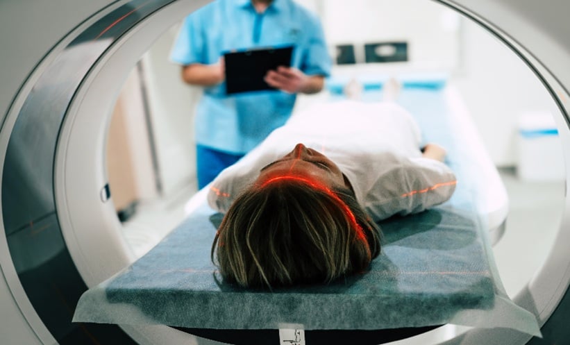
MRI-DERIVED RADIOMICS may help clinicians identify high-risk tumour deposits in rectal cancer before surgery, according to a new dual-centre study suggesting artificial intelligence could enhance treatment planning and outperform conventional image assessment.
Dual-Centre Study Evaluates MRI-Based Radiomics Models
The retrospective analysis evaluated 729 patients with rectal cancer treated between 2018 and 2024, with 376 ultimately included for model development and validation. Researchers investigated whether radiomics features extracted from MRI scans could stratify patients based on tumour deposit (TD) burden, an important prognostic factor associated with disease progression and poorer outcomes.
Participants were categorised into three groups according to TD number: no deposits, one to two deposits, and three or more deposits. Using machine learning, investigators built predictive models based on features derived from the primary tumour, the largest mesorectal nodule, and a fusion approach combining both datasets. The models were trained using the XGBoost algorithm and assessed across multiple performance metrics, including area under the curve (AUC), accuracy, precision, recall, and F1 score.
Among the approaches tested, the fusion radiomics model demonstrated the strongest performance. It achieved AUC values of 0.873 in the test set and 0.858 in the validation cohort, alongside accuracy rates approaching 80%. Notably, this combined model outperformed two experienced radiologists, whose accuracy ranged from 0.589 to 0.676, highlighting the potential clinical value of quantitative image analysis.
The tumour-only model also showed robust predictive ability, with AUCs exceeding 0.84, while the nodule-based model achieved slightly lower but still clinically relevant performance. Researchers suggest that integrating tumour characteristics with features of the largest mesorectal nodule provides a more comprehensive representation of disease biology, improving classification of TD burden.
Implications for Personalised Treatment Planning
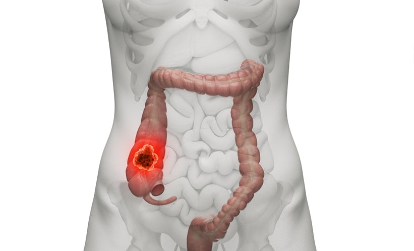
Tumour deposits are recognised as an adverse prognostic marker in rectal cancer, influencing staging and therapeutic decisions. However, accurately identifying TD before surgery remains challenging using standard imaging interpretation alone. The authors propose that MRI-based radiomics could offer an objective tool to support preoperative risk stratification, potentially guiding personalised treatment strategies such as intensified neoadjuvant therapy or closer surveillance.
Although promising, the study’s retrospective design and limited external validation highlight the need for prospective multicentre research before widespread clinical adoption. Nonetheless, the findings add to growing evidence that AI-driven radiomics may augment radiologist performance and refine precision oncology pathways in colorectal cancer care.
Reference
Zhang C et al. MRI-derived radiomics for risk stratification of tumour deposits in rectal cancer: a dual-centre study. Insights Imaging. 2026;17:31.
- Author: