Abstract
Mixed connective tissue disease (MCTD), an overlap syndrome described by Gordon Sharp in 1972, combines clinical features of systemic lupus erythematosus, systemic sclerosis, and polymyositis. Though renal involvement is relatively uncommon, severe thrombocytopenia, pulmonary arterial hypertension, and polyserositis as initial manifestations remain rare clinical presentations. This case series highlights unusual manifestations of MCTD.
Case 1 describes a pregnant woman presenting with severe immune-mediated thrombocytopenia, successfully managed with immunosuppressive therapy. Case 2 highlights pulmonary arterial hypertension presenting with progressive dyspnea and respiratory symptoms, treated with sildenafil and immunosuppressive therapy. Case 3 involves polyserositis with ascites mimicking chronic liver disease, diagnosed as MCTD after exclusion of infectious, hepatic, and malignant etiologies.
All patients demonstrated positive antinuclear antibodies with anti-U1 ribonucleoprotein (anti-U1 RNP) antibodies, fulfilling key serological criteria for MCTD. These cases emphasize the heterogeneous clinical spectrum of anti-RNP associated disease and highlight the importance of considering MCTD in atypical multisystem presentations. Early diagnosis, multidisciplinary management, and individualized immunosuppressive therapy are essential to optimize outcomes.
Key Points:
1. Anti-U1 ribonucleoprotein associated disease may present with incomplete or evolving clinical features.
2. Severe thrombocytopenia and polyserositis may represent lupus-like manifestations of mixed connective tissue disease and may mimic infection or chronic liver disease.
3. Pulmonary arterial hypertension is an important complication requiring early screening with early recognition and multidisciplinary management to improve clinical outcomes.
INTRODUCTION
Mixed connective tissue disease (MCTD), first described by Gordon Sharp in 1972, is a systemic autoimmune overlap syndrome characterized by clinical features of systemic lupus erythematosus (SLE), systemic sclerosis, and polymyositis.1 Common manifestations include polyarthritis, Raynaud’s phenomenon, sclerodactyly, esophageal dysmotility, pulmonary fibrosis, and inflammatory myopathy.
Although renal involvement is less frequent, occurring in approximately 25% of cases, hematological manifestations such as severe immune-mediated thrombocytopenia as an initial presentation are uncommon. Laboratory findings typically include high-titer antinuclear antibodies (ANA) with antibodies directed against U1-ribonucleoprotein (U1-RNP), which play an important role in disease classification and prognostication.
According to the Alarcón-Segovia diagnostic criteria, the diagnosis of MCTD requires high-titer anti-U1 RNP antibodies along with at least three clinical features including Raynaud phenomenon, synovitis, myositis, swollen hands, or acrosclerosis. However, it is well recognized that patients may initially present with incomplete or evolving clinical features, making early diagnosis challenging.2
Treatment strategies are largely extrapolated from therapies used in related connective tissue diseases and include corticosteroids, hydroxychloroquine (HCQ), and immunosuppressive agents such as azathioprine, cyclophosphamide, and mycophenolate mofetil.
The authors present three cases of MCTD with atypical presentations, including immune thrombocytopenia during pregnancy, pulmonary arterial hypertension (PAH), and polyserositis presenting as ascites, highlighting diagnostic challenges and therapeutic considerations.
CASE PRESENTATIONS
Case 1: Immune Thrombocytopenia as Initial Manifestation of MCTD
A 26-year-old pregnant woman at 4 months gestation presented with rash, hematuria, gum bleeding, and epistaxis for 3 days. She also reported fever, vomiting, headache, generalized weakness, and dizziness. She denied history of arthralgia, myalgia, dyspnea, palpitations, abdominal pain, or neurological symptoms.
Clinical examination revealed severe pallor without icterus or lymphadenopathy. Laboratory evaluation demonstrated severe thrombocytopenia (5,000 /mm3) with normal platelet morphology and normocytic normochromic anemia (hemoglobin: 9.5 g/dL). Liver function tests, kidney function tests, and urine examination were normal.
Rapid screening test for HIV 1 and 2 was negative. Two units of random donor platelets were transfused, resulting in a transient rise in platelet count to 14,000 /mm3. The patient developed erythematous rash and pruritus following transfusion.
Past history revealed a similar episode during her previous pregnancy associated with bleeding manifestations requiring transfusion support.
Autoimmune evaluation showed ANA positivity with homogeneous pattern (1:320), elevated erythrocyte sedimentation rate (21 mm/hr), and elevated C-reactive protein (48 mg/L). U1-RNP/Sm IgG antibodies were positive. Anti-dsDNA, anti-SSA, anti-SSB, anti-Scl-70, anti-histone antibodies, and antiphospholipid antibodies were negative. Complement levels (C3 and C4) were normal.
Bone marrow examination demonstrated megakaryocytic thrombocytopenia suggestive of peripheral immune destruction.
Severe thrombocytopenia in MCTD is considered a lupus-like manifestation and may occur due to immune-mediated platelet destruction, similar to immune thrombocytopenic purpura. Although antiphospholipid antibodies may be associated with cytopenias, they were negative in this patient.
Based on clinical and serological findings, a diagnosis of MCTD presenting with immune thrombocytopenia was made. The patient was treated with intravenous methylprednisolone, HCQ, and azathioprine with good clinical response.
Case 2: Pulmonary Arterial Hypertension in Anti-RNP Associated Disease
A 42-year-old female presented with productive cough, breathlessness, chest pain, and palpitations for 2 months. She also reported lower limb pain, painful ulcers over the anterior tibial shin region, and oral mucocutaneous painful ulcers. Hemoglobin was 9 g/dL and platelet count was normal. Liver enzymes were elevated (aspartate aminotransferase: 1,289 U/L; alanine aminotransferase: 294 U/L). CRP was elevated (126 mg/dL).
Tuberculosis work-up, including acid-fast bacillus smear and GeneXpert® (Danaher Corporation, Washington, D.C., USA), was negative.
High-resolution CT (HRCT) thorax demonstrated cardiomegaly, fibrotic lung changes, traction bronchiectasis, and PAH. Echocardiography revealed severe tricuspid regurgitation and severe PAH with dilated inferior vena cava.
Right ventricular systolic pressure of 78 mmHg estimated on echocardiography suggested severe pulmonary hypertension. Although right heart catheterization remains the gold standard for diagnosis, it was not performed due to clinical considerations and resource limitations. Autoimmune profile demonstrated ANA positivity with speckled pattern and positive anti-U1 RNP antibodies.
PAH is a recognized complication of anti-RNP associated disease and may occur due to immune mediated endothelial dysfunction, leading to progressive increase in pulmonary vascular resistance and right heart failure.
The patient was treated with sildenafil, azathioprine, corticosteroids, and antibiotics, resulting in symptomatic improvement.
Case 3: Polyserositis Presenting as Ascites Mimicking Chronic Liver Disease
A 30-year-old female presented with abdominal distension, dyspnea on exertion, and polyarthralgia for 3 months.
Examination revealed generalized edema and ascites. Laboratory investigations showed hemoglobin of 9.8 g/dL with normal liver and renal parameters. Ascitic fluid analysis revealed lymphocytic predominance with low adenosine deaminase (ADA; 4.4 U/L), making tuberculosis unlikely.
HRCT thorax demonstrated pericardial effusion, mild pleural effusion, and pulmonary inflammatory changes including tree-in-bud appearance and ground-glass opacities.
Given prior history of tuberculosis, extensive evaluation including sputum acid-fast bacilli, GeneXpert, Mantoux test, and ascitic fluid ADA was performed to exclude active infection.
Autoimmune profile revealed ANA positivity with fine speckled pattern and positive anti-U1 RNP antibodies.
Evaluation for autoimmune liver disease was performed and revealed negative anti-mitochondrial and anti-smooth muscle antibodies. Viral hepatitis serology including hepatitis B surface antigen and anti-hepatitis C virus were also negative.
Polyserositis in MCTD may result from immune complex mediated inflammation involving pleura, pericardium, and peritoneum.
The patient improved with corticosteroids, HCQ, and diuretics.
DISCUSSION
MCTD is a systemic autoimmune disorder predominantly affecting women, with a reported female-to-male ratio of approximately 3:1. The clinical spectrum of MCTD is notably diverse, ranging from mild symptoms such as arthralgia to severe complications, including PAH, interstitial lung disease, and polyserositis.3 The incidence of MCTD remains relatively low, with studies estimating around 2.1 cases per million per year. Despite its rarity, the disease is of particular interest due to its overlap features with SLE, scleroderma, and polymyositis, along with its characteristic serological marker, anti-U1 RNP antibodies.4
Several cases have been documented, highlighting rare presentations of MCTD (Table 1).5-9
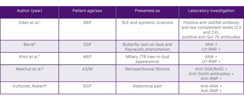
Table 1: MCTD with rare presentation.5-9
ANA: antinuclear antibodies; anti-dsDNA: anti-double-stranded DNA; F: female; M: male; MCTD: mixed connective tissue disease; RNP: ribonucleoprotein; SLE: systemic lupus erythematosus; TB: tuberculosis.
Thrombocytopenia as an Initial Manifestation
Thrombocytopenia, as seen in Case 1, is a rare presentation of MCTD. This condition typically results from peripheral destruction or immune-mediated mechanisms. Although gestational thrombocytopenia is common and usually benign, severe thrombocytopenia necessitates thorough investigation for underlying autoimmune or immune-mediated causes.10 In the context of MCTD, its occurrence during pregnancy is particularly rare. This underscores the importance of considering autoimmune connective tissue diseases in the differential diagnosis of unexplained thrombocytopenia in pregnant patients. Early diagnosis and timely initiation of immunosuppressive therapy are essential to prevent maternal and fetal complications.11
Management of MCTD during pregnancy poses significant challenges due to potential risks to both mother and fetus. Corticosteroids remain the mainstay of treatment for controlling disease activity. HCQ is considered safe and is often used to manage autoimmune conditions during pregnancy. Azathioprine, an immunosuppressive agent, is also deemed relatively safe for use during pregnancy when necessary.12 Close monitoring of maternal and fetal health is imperative, with adjustments to therapy made based on disease activity and gestational age.13 Studies suggest that active disease at conception and during pregnancy may increase the risk of adverse outcomes, highlighting the importance of achieving disease remission before conception when possible. Additionally, regular monitoring for potential complications such as preeclampsia, intrauterine growth restriction, and preterm birth is recommended.13
Managing MCTD during pregnancy presents unique challenges. The primary goal is to control disease activity while minimizing risks to both mother and fetus. Corticosteroids are the mainstay of therapy, often used in conjunction with HCQ, which is considered safe during pregnancy. Azathioprine, another immunosuppressive agent, is relatively safe when deemed necessary.
Close monitoring of maternal and fetal health is crucial, with therapy adjustments based on disease activity and gestational age. A multidisciplinary approach, involving rheumatologists, obstetricians, and neonatologists, is recommended to optimize outcomes. Studies emphasize that achieving disease remission before conception significantly reduces the risk of adverse pregnancy outcomes. Regular monitoring for complications such as preeclampsia, intrauterine growth restriction, and preterm birth is essential.13
Pulmonary Manifestations and PAH
Pulmonary involvement, particularly PAH, is a serious complication of MCTD, as illustrated in Case 2. PAH arises from immune-mediated vasculopathy and is associated with significant morbidity and mortality. Studies report the prevalence of PAH in MCTD patients to be between 7–23%. The condition is characterized by increased pulmonary vascular resistance, eventually leading to right heart failure.14,15
In this case, the patient presented with respiratory symptoms, chest pain, and palpitations, with PAH confirmed via HRCT and echocardiography. Management included sildenafil, a phosphodiesterase-5 inhibitor, to improve pulmonary hemodynamics, along with immunosuppressive therapy comprising corticosteroids and azathioprine. Broad-spectrum antibiotics were administered to mitigate potential secondary infections during immunosuppression.16
Recent studies highlight the importance of early diagnosis and aggressive management of PAH in MCTD. Early intervention with PAH-specific therapies, combined with immunosuppressants, can improve survival rates. A multidisciplinary team approach is critical for effective management, underscoring the need for vigilance in identifying early signs of PAH in patients with MCTD.17
Polyserositis and Ascites asInitial Presentation
Polyserositis, a rare but recognized manifestation of MCTD, presents diagnostic challenges due to its overlap with other conditions such as chronic liver disease. In Case 3, the patient presented with ascites, joint pain, and shortness of breath, leading to an extensive diagnostic workup. The presence of pericardial and pleural effusions, along with pulmonary hypertension, suggested MCTD, confirmed by serological markers such as anti-U1 RNP antibodies.18
Differentiating MCTD from chronic liver disease required careful evaluation. Normal liver function tests, low ascitic fluid ADA levels, and negative tuberculosis markers excluded hepatic or infectious causes.19 Immunosuppressive therapy, including corticosteroids and HCQ, was initiated to control systemic inflammation, while diuretics were used to manage fluid accumulation.20
Anti-U1 RNP antibodies are strongly associated with vascular dysfunction, immune complex deposition, and cytokine mediated inflammation contributing to diverse manifestations including cytopenias, pulmonary hypertension, and serositis.
Table 1 highlights previously reported rare manifestations of MCTD. The present case series adds to existing literature by demonstrating immune thrombocytopenia during pregnancy, severe pulmonary hypertension, and polyserositis presenting as ascites, thereby expanding the clinical spectrum of anti-RNP associated disease.
Thrombocytopenia
Immune thrombocytopenia is an uncommon initial manifestation of MCTD but is well described in lupus spectrum disorders. Mechanisms include immune-mediated platelet destruction and antibody-mediated peripheral consumption.
Pulmonary Hypertension
PAH occurs in 7–23% of patients with MCTD and represents a major cause of morbidity. Anti-RNP positivity is associated with increased risk of pulmonary vascular disease and interstitial lung disease.
Polyserositis
Serositis involving pleural, pericardial, and peritoneal surfaces may mimic chronic liver disease or infection. Careful exclusion of tuberculosis, malignancy, and hepatic disease is essential before attributing ascites to autoimmune etiology.
Anti-Smith antibodies are specific for SLE, and may rarely coexist with anti-U1 RNP in overlap connective tissue disease. Diagnosis should rely on clinical correlation, not antibody alone. Absence of classical SLE features favors MCTD spectrum rather than pure SLE.
According to the Alarcón-Segovia criteria, classification of MCTD requires high-titer anti-U1 RNP antibodies along with at least three characteristic clinical features such as Raynaud’s phenomenon, swollen hands, synovitis, myositis, or acrosclerosis. None of the three patients in the present series completely fulfilled classical Alarcón-Segovia criteria at presentation. However, all demonstrated strong anti-U1 RNP positivity with compatible multisystem autoimmune manifestations suggestive of anti-RNP associated overlap connective tissue disease.
Case 1 was interpreted as an evolving/incomplete MCTD phenotype, with anti-U1 RNP positivity and severe immune thrombocytopenia representing a lupus-like hematological manifestation within the anti-RNP disease spectrum. It was therefore interpreted as an early evolvingMCTD phenotype.
Case 2 similarly represented an evolving anti-RNP associated overlap phenotype. Although classical criteria were not fully satisfied, the presence of PAH and interstitial pulmonary involvement in association with anti-U1 RNP antibodies strongly supported evolving MCTD spectrum disease.
Case 3 was also considered an evolving/incomplete MCTD phenotype, characterized by anti-U1 RNP positivity with polyserositis involving pleural, pericardial, andperitoneal surfaces.
These cases illustrate that anti-U1 RNP associated disease may initially manifest with incomplete or atypical overlap features before evolving into classical MCTD on longitudinal follow-up.
Treatment Considerations
Management of MCTD is individualized based on organ involvement and often follows treatment strategies used in related connective tissue diseases. Corticosteroids remain first-line therapy for inflammatory manifestations. HCQ is commonly used for long-term disease control and prevention of disease flares. Azathioprine acts as steroid sparing immunosuppressive therapy. Sildenafil improves pulmonary hemodynamics in patients with PAH. Multidisciplinary management involving rheumatologists, pulmonologists, cardiologists, and obstetricians is essential for optimal outcomes.
CONCLUSION
MCTD demonstrates a broad clinical spectrum and may initially present with atypical manifestations, leading to diagnostic delay.
Further research is required to improve risk stratification and optimize therapeutic strategies in patients with anti-RNP associated autoimmune disease.