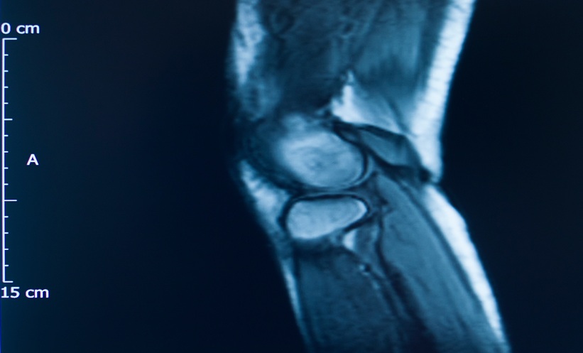
QUANTIFYING joint inflammation scoring rapidly on extremity magnetic resonance imaging scans has approached expert reader performance using deep learning. Investigators developed and validated the Automatic Deep Learning-Based MRI Analysis of Inflammatory Signs in Rheumatoid Arthritis system, termed ADMIRA. Evaluated across 2,254 subjects from four clinical cohorts, the neural network estimated inflammatory features on fat-saturated, contrast-enhanced T1-weighted scans of the wrists, metacarpophalangeal joints, and metatarsophalangeal joints. Manual evaluation using the Rheumatoid Arthritis Magnetic Resonance Imaging Scoring framework requires extensive time and faces notable inter-reader variability. By contrast, the automated system delivers assessments in less than one second per input volume on standard computing infrastructure.
Deep Learning Achieves Expert-Level Joint Inflammation Scoring
The deep learning pipeline processes three-dimensional magnetic resonance volumes by executing automated background clearance and selecting the seven most informative slices from transversal and coronal acquisitions. Multi-view cross-attention modules merge spatial data from both planes to replicate the multiplanar assessment performed by clinical experts during visual evaluation.
On hold-out test sets, the algorithm demonstrated correlation coefficients and intraclass correlation coefficients approaching 0.9 for tenosynovitis and synovitis. The system evaluated both total anatomical region burden and individual structural components. Validation against an independent trial cohort confirmed intraclass correlation coefficients near 0.8 for soft-tissue inflammation. Agreement for bone marrow edema proved lower, achieving values of 0.8 on test sets and 0.7 on independent validation. The investigators observed that less distinct signal margins and skewed severity distributions in bone lesions contributed to this performance difference.
Clinical Utility and Workflow Integration
The platform provides two flexible deployment routes for rheumatology and radiology practices. The first pathway calculates a composite regional inflammation score directly, maximizing computational speed for high-volume triage. The second pathway determines severity across specific anatomical sites, such as the extensor carpi ulnaris tendon sheath, before calculating overall totals.
To verify clinical interpretability, researchers applied rescaled regression activation mapping. The generated heatmaps confirmed that model predictions localized to true areas of synovial proliferation and peritendinous effusion rather than imaging artifacts. This end-to-end regression method eliminates manual segmentation steps, offering reproducible, high-throughput disease monitoring to support early intervention pathways.
Reference
Li Y et al. Automatic joint inflammation estimation based on regression neural networks. Med Phys. 2025;52:e70010.