Funding Statement: This symposium was funded by Insmed Incorporated. The publication of this article was sponsored by Insmed Incorporated.
Chairperson: Oriol Sibila1
Speakers: Merete Long,2 Pieter Goeminne,3 Diego J. Maselli,4 Deval Gor,5 Eva Polverino6
1. Hospital Clinic de Barcelona, Spain
2. University of Dundee, UK
3. VITAZ Hospitals Sint-Niklaas, Belgium
4. UT Health San Antonio, Texas, USA
5. Insmed Incorporated, Bridgewater, New Jersey, USA
6. Respiratory Disease Department, Hospital Universitari Vall d’Hebron, Vall d’Hebron Institut de Recerca (VHIR), Vall d’Hebron Barcelona Hospital Campus, Spain
Conflicts of interest: Sibila has received speaker and consultancy fees from Boehringer Ingelheim and Insmed. Long has received speaker fees from Insmed. Goeminne has received fees for lectures and participation on advisory boards and data monitoring committees, and support to attend meetings, from Insmed, Boehringer Ingelheim, MSD, Pfizer, GSK, and AstraZeneca. Maselli has received speaker and consultancy fees from AstraZeneca, GSK, Amgen, Sanofi/Regeneron, and Insmed; and research funding from the National Institutes of Health, the Bill & Melinda Gates Foundation, Seastar, the COPD Foundation, Boehringer Ingelheim, and Chiesi. Gor is an employee and shareholder of Insmed Incorporated. Polverino has received grants from Grifols; and speaker and consultancy fees from Chiesi, CSL Behring, Grifols, GSK, Insmed Incorporated, Moderna, Pfizer, Boehringer Ingelheim, and Pari.
Acknowledgements: Medical writing assistance was provided by Christos Evangelou, MedRight Medical Writing & Editing, San Diego, California, USA.
Disclaimer: The symposium content and views expressed herein are those of the speakers and not necessarily of the sponsor. Insmed Incorporated participated in the study design, research, analysis, data collection, and interpretation of data, as well as review and approval of this article.
Keywords: Bronchiectasis, bronchiectasis diagnosis, diagnostic delay, exacerbations, healthcare resource utilisation, neutrophil elastase, neutrophilic inflammation, Pseudomonas aeruginosa, psychosocial impact, quality of life.
Citation:EMJ Respir. 2026;14[1]:49-57. https://doi.org/10.33590/emjrespir/I56WH8B4
Meeting Summary:
Speakers at a symposium during the European Respiratory Society (ERS) Congress 2026 in Barcelona, Spain, discussed the burden of bronchiectasis, delayed diagnosis, and underlying inflammatory mechanisms. Bronchiectasis is a chronic, neutrophil-driven inflammatory airway disease characterised by progressive structural lung damage that is visualised as distinct radiographic changes on CT scan. Bronchiectasis is increasingly prevalent and underdiagnosed. The diagnosis of bronchiectasis is often delayed, with reported delays of 3.5–17.0 years from symptom onset to diagnosis. Factors contributing to diagnostic delays include symptom overlap with asthma and COPD, low disease awareness, and underutilisation of CT imaging. Speakers described the pathophysiology of bronchiectasis as a self-perpetuating cycle of airway inflammation, chronic infection, impaired mucociliary clearance, and structural lung damage. Two posters presented at the ERS Congress 2026 highlighted data on the real-world and patient-reported impact of exacerbations in bronchiectasis. Data from the BURDEN study, which surveyed 713 people with bronchiectasis across five European countries, showed that exacerbations had an additional negative impact on sleep, self-care, daily tasks, emotional wellbeing, family life, social life, and work, with the impact intensifying with exacerbation frequency. A separate analysis of the Spanish BIG-PAC electronic medical record database showed that more frequent or severe exacerbations were associated with greater use of antibiotics, inhaled corticosteroids, and bronchodilators, and more outpatient visits and hospitalisation. Together, these findings support the importance of earlier recognition and more proactive management of bronchiectasis.
Introduction: Epidemiology and Unmet Needs
Bronchiectasis has historically been regarded as a rare, ‘orphan’ condition, but the data presented at the symposium indicate that its prevalence is increasing.1 Global prevalence of bronchiectasis, presented by Oriol Sibila, Hospital Clinic de Barcelona, Spain, at the symposium, ranges from approximately 50 to more than 1,000 cases per 100,000 population.2,3 In the USA, the prevalence of bronchiectasis rose from 52.3 per 100,000 to 714.0 per 100,000 between 1999–2014, and the UK has reported an annual rise in recorded cases of between 8–20%.2 The prevalence of bronchiectasis increases with age, with a mean age of diagnosis of approximately 67 years.2 Analysis of the European Bronchiectasis registry (EMBARC) data (N=16,963) showed that 60.9% of patients with bronchiectasis were women.2,4
According to data presented at the symposium, the diagnosis of bronchiectasis is frequently established at an advanced stage of disease progression, reflecting a pattern of diagnostic delay.1 A Spanish national registry study reported a median diagnostic delay of 12 years from symptom onset to diagnosis, which was approximately 2.1 years longer in women than in men.5 A UK specialist-clinic cohort showed a 17-year diagnostic delay, while an Italian multicentre observational point-prevalence study reported a delay of approximately 3.5 years between symptom onset and diagnosis.6,7 The speakers pointed out that the prolonged diagnostic delay represents a missed opportunity for intervention, potentially leading to disease progression.
The speakers at the symposium discussed the clinical burden of bronchiectasis.1 Approximately 35% of patients in EMBARC had severe disease at registry entry, and 36% had airflow obstruction, with forced expiratory volume in 1 second (FEV1) below 30% predicted in 3.6% and FEV1 30–49% predicted in 13.8% of patients.4 Among patients in the EMBARC study, 26.4% had been hospitalised for an exacerbation in the preceding year, and the median number of exacerbations per patient was two (interquartile range: 1–4).4 Patient mortality over long-term follow-up (≥13 years) across studies has been reported to be 29.7–36.0%.2
The speakers also highlighted the substantial economic burden of bronchiectasis, with total annual healthcare costs in the UK and Spain estimated at 448 million USD.8,9 The vicious cycle of infection, decline, and cost in bronchiectasis raises the risk of poor quality of life and independently increases future mortality risk.1,10,11
The speakers concluded that earlier diagnosis and more effective early treatment are needed to reduce the clinical and economic burden of bronchiectasis by reducing exacerbations and preserving lung function.1
Understanding the Underlying Disease Process
Merete Long, University of Dundee, UK; and Pieter Goeminne, VITAZ Hospitals Sint-Niklaas, Belgium, discussed the current understanding of bronchiectasis pathophysiology.1 The 2025 ERS clinical practice guideline describes the pathophysiology of bronchiectasis as a vicious vortex, in which chronic airway inflammation, chronic and recurrent airway infection, impaired mucociliary clearance, and progressive structural lung damage are all interlinked and amplify one another (Figure 1).12,13 Chronic inflammation is an underlying driver of disease, resulting from dysregulated neutrophilic activity dependent on activation of neutrophil serine proteases by an upstream enzymatic step during neutrophil maturation, generating an imbalance between proteases and antiproteases in the airway.12
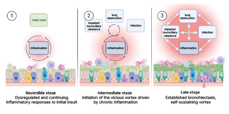
Figure 1: Inflammation as a driver of bronchiectasis initiation and disease progression.
Reprinted from Long MB et al.13 © 2024, with permission from Elsevier, Amsterdam, the Netherlands.
Neutrophil elastase was discussed as a marker that has been associated with disease activity and may serve as a measurable indicator of underlying pathological processes.1 Higher activity of sputum neutrophil elastase has been associated with higher all-cause mortality over 3 years, shorter time to next hospitalisation for severe exacerbation, higher bacterial load, and shorter time to next exacerbation (overall and severe exacerbation).14 Goeminne presented data illustrating how this inflammatory activity translates into impaired mucociliary clearance at a tissue level.1 Neutrophil elastase has been associated with increased sputum volume and induces goblet cell hyperplasia leading to increased mucus production.15
Azurocidin-1 was also presented as a mediator of airway pathology in bronchiectasis.1 Analysis of azurocidin-1 levels in the sputum of 197 patients with bronchiectasis showed that high azurocidin-1 levels were associated with greater disease severity, worse lung function, and increased frequency of exacerbations.16 In in vitro experiments, the addition of azurocidin-1 to respiratory epithelial cells reduced ciliary beat frequency by 2.988 Hz and increased the epithelial disruption score.16
The speakers noted that, collectively, these findings suggest that the inflammatory, infective, mucociliary-clearance, and structural-damage pillars of the vicious vortex are mechanistically interconnected, such that dysregulated activity in one pillar can compound the others.1 They also highlighted that this interdependence is central to why timely diagnosis and early intervention are essential, as initiating effective management before the vortex accelerates may help interrupt the cycle of tissue damage.1
Why the Diagnosis is Delayed or Missed, and Impact of Delays
At the symposium, Diego J. Maselli, UT Health San Antonio, Texas, USA, addressed why bronchiectasis is often overlooked or misattributed to other respiratory conditions.1
Bronchiectasis shares its cardinal symptoms with asthma and COPD, which can make it difficult to distinguish.1 Chronic productive cough, purulent or mucopurulent sputum, and recurrent exacerbations and respiratory infections are common to bronchiectasis;17 asthma (which also features variable wheeze, dyspnoea, cough, airflow obstruction, and trigger-related symptoms such as cold, virus, or exercise);18 and COPD (which also features progressive dyspnoea with chronic cough, sputum production, and a history of recurrent lower respiratory tract infections or risk-factor exposure).19 Because these symptoms overlap, airway dilation on CT is a key distinguishing hallmark feature that clinicians should look for to help identify bronchiectasis.17
Bronchiectasis frequently coexists with asthma and COPD.1 An Italian cohort study of 232 patients with bronchiectasis showed that 26.7% and 18.5% of patients had coexisting asthma and COPD, respectively.7 Among the 16,963 patients with bronchiectasis in the EMBARC study, 5,267 (31.0%) had concomitant asthma.20 In addition, 4,324 of 16,963 patients with bronchiectasis in the EMBARC study also had a diagnosis of COPD.21
The patient journey data presented at the symposium indicate that delays exist at multiple points in the diagnostic journey.1,7,22 Potential drivers of delay and underdiagnosis include low awareness of bronchiectasis among clinicians and patients; overlapping symptoms with asthma, COPD, and chronic bronchitis; misdiagnosis, with 25.7% of patients in the EMBARC/European Lung Foundation study reported as previously misdiagnosed, most often with asthma, then chronic bronchitis and COPD; the limited sensitivity of chest radiography and symptoms alone; and access barriers, with 50.2% of patients in the EMBARC/European Lung Foundation study finding primary care access during an exacerbation difficult.7,22
The symposium also reported data on the clinical impact of diagnostic delays in patients with bronchiectasis.1 Analysis of multicentre EMBARC study data showed that longer symptom duration before diagnosis (analysed per additional decade) was associated with increased risk of Pseudomonas aeruginosa infection, a higher exacerbation rate, increased odds of hospitalisation, and reduced lung function at presentation.23 The analysis also showed that the onset of bronchiectasis symptoms follows a bimodal age distribution, with one peak in childhood or early adulthood and a larger peak around the sixth and seventh decades, and that earlier onset, corresponding to longer symptom duration by the time of assessment, is associated with lower FEV1 % predicted.23 These findings led the speakers to conclude that early recognition and diagnosis of bronchiectasis is essential to help reduce future disease burden.1
When to Suspect Bronchiectasis, and How Overlap is Defined
Maselli’s session at the symposium included discussion of cues for raising clinical suspicion.1 Clinical features that should raise clinical suspicion for bronchiectasis include chronic productive cough with daily mucopurulent or purulent sputum; recurrent lower respiratory tract infections or exacerbations; unexplained haemoptysis; asthma or COPD responding poorly to treatment or lacking a clear atopic or smoking basis; and isolation of P. aeruginosa or non-tuberculous mycobacteria from sputum.24
Maselli outlined criteria for defining the overlap between bronchiectasis and COPD.1 The ROSE criteria, which were developed through a Delphi consensus process by the EMBARC Airways Working Group, provided a structured definition of bronchiectasis–COPD overlap syndrome based on radiological findings (bronchiectasis on CT), obstruction (FEV1/forced vital capacity below 0.7), symptoms, and exposure (10 or more pack-years of smoking).25 Applying the ROSE criteria to the EMBARC cohort revealed overdiagnosis of COPD in patients with bronchiectasis: 22.2% of patients labelled as having COPD had no airflow obstruction, and 31.9% lacked a significant smoking history.21
Asthma also commonly co-occurs with bronchiectasis; concomitant asthma was present in an estimated 31.0% of the EMBARC cohort and more frequently affected patients who were younger, female, never-smokers, and of higher BMI than those with bronchiectasis alone.20 Patients with concomitant asthma in the EMBARC study also showed higher rates of rhinosinusitis, nasal polyps, eosinophilia, and Aspergillus sensitisation, and were more likely to experience three or more exacerbations than patients without concomitant asthma.20
Exacerbations: Definition, Trajectory, and Impact on Survival
Maselli discussed how exacerbations are defined, how they behave over time, and their impact on patient survival.1
The 2025 ERS clinical practice guideline defines an exacerbation as a worsening of symptoms that exceeds day-to-day variability and requires a change in management.13 Common symptoms of exacerbation include cough, a change in sputum volume or consistency, sputum purulence, dyspnoea or reduced exercise tolerance, fatigue or malaise, and haemoptysis, alongside clinical features such as fever, wheeze, general discomfort, anorexia, weight loss, pleuritic chest pain, and changes on chest examination.12,26,27
A severe exacerbation is defined as an exacerbation that requires hospitalisation or intravenous antibiotics.12 Symptoms of severe exacerbations include tachypnoea, acute or acute-on-chronic respiratory failure, a significant fall in oxygen saturation or respiratory function, hypercapnia, haemoptysis, new cyanosis, new signs of cor pulmonale, haemodynamic instability, or impaired cognitive function.12
Maselli presented data on exacerbation trajectory that suggest that exacerbation frequency in a given year is associated with mortality.1 A multicentre study of patients with bronchiectasis followed for up to 5 years showed that patients with more exacerbations in an initial year tended to have more in the 2 subsequent years.10 Frequent exacerbations were associated with high disease severity, worse health status, worse quality of life, and increased mortality during follow-up.10 Analysis of the EMBARC study data showed that a higher number of exacerbations in the previous year was associated with an increased risk of exacerbations during follow-up.28 A different analysis of data from 92,529 patients with bronchiectasis demonstrated differences in mortality based on exacerbation frequency.29 By the end of the 5-year follow-up, survival rates were 65.4% in patients with no exacerbations at baseline, 62.6% in patients with one exacerbation, and 55.3% in those with two or more exacerbations.29 Median time to death was 70.4 months for patients with two or more exacerbations at baseline and was not reached in patients with one or no exacerbations at baseline.29
The Psychosocial Burden of Exacerbations: Findings from the BURDEN Study
Study Objective, Design, and Demographics
Exacerbations in bronchiectasis have been associated with poorer health-related quality of life.11 In a poster presented at the ERS Congress 2026, Eva Polverino, Hospital Universitari Vall d’Hebron, Vall d’Hebron Institut de Recerca (VHIR), Vall d’Hebron Barcelona Hospital Campus, Spain, and colleagues reported data on the patient-reported psychosocial impact of exacerbations on daily life.30 The presented data were part of the BURDEN study, which aimed to assess the psychosocial impact of exacerbations from the perspectives of people living with bronchiectasis in Europe.30
The investigators used qualitative interviews conducted in the USA and Europe and a quantitative online survey, co-created with HCPs and patient experts.30 The poster reported quantitative results from the European arm, which included 713 respondents with a bronchiectasis diagnosis (excluding cystic fibrosis), ≥1 exacerbation in the previous year, ≥18 years, and living in France, Germany, Italy, Spain, or the UK.30 Respondents were predominantly older (74.1% aged 60 years or above) and female (82.6%), and most (56.0%) had been diagnosed 5 or more years earlier.30 The most commonly reported comorbidities were asthma (32.5%), gastro-oesophageal reflux disease (25.7%), hypertension (23.4%), COPD (21.5%), bone and joint disease (20.9%), chronic sinusitis (19.1%), and non-tuberculous mycobacterial lung disease (12.1%).30
Frequency and Psychosocial Impact of Exacerbations
Over the previous 12 months, 26.2% of respondents had experienced one exacerbation, and 73.8% had experienced ≥2 exacerbations (27.1% with two and 46.7% with ≥3).30 The proportion of respondents reporting ≥3 exacerbations ranged from 34.9% in Italy to 39.4% in Spain, 51.5% in the UK, 57.8% in France, and 58.8% in Germany.30
Participants rated the additional negative impact of exacerbations, versus periods without exacerbations, across seven dimensions of daily life (sleep quality, self-care activities, daily tasks, emotional wellbeing, family life, social life, and work or study) on a scale from 1 (indicating no additional negative impact at all) to 10 (indicating an extreme amount of additional negative impact).30
Exacerbations were associated with additional negative impact across all dimensions of daily living, with effects increasing with exacerbation frequency; differences between the one-exacerbation and three-or-more-exacerbation groups were statistically significant across all dimensions except work or study.30 For respondents with two exacerbations in the previous 12 months, mean scores ranged from 3.9 (self-care activities) to 5.9 (emotional wellbeing); for those with three or more exacerbations, scores ranged from 4.2 (self-care activities) up to 6.4 (emotional wellbeing) and 6.5 (social life).30
Separately, 73.1% of the 408 respondents asked this question reported that they felt every exacerbation worsened their overall condition.30
Limitations
Study limitations include that diagnoses and exacerbations were self-reported without medical record verification, that participants may have been subject to recall bias, and that some may have found it difficult to distinguish exacerbation from stable-state experience.30
Conclusion
The authors of the poster concluded that exacerbations have a substantial impact on people with bronchiectasis in Europe across psychosocial aspects and dimensions of daily living.29 These data highlight the importance of HCPs addressing patient-reported outcomes and optimising exacerbation management.30
Treatment Burden and Healthcare Resource Use: Real-World Data from Spain
Study Objective, Design, and Population
The poster presented at the ERS Congress 2026 by Deval Gor, Insmed Incorporated, Bridgewater, New Jersey, USA, and colleagues used the Spanish BIG-PAC electronic medical record database to characterise treatment patterns and healthcare resource utilisation among adults with bronchiectasis (excluding cystic fibrosis), stratified by exacerbation frequency, in two cohorts: a 2019 cohort (N=4,469) assessing treatment use, and a 2023 cohort (N=5,671) assessing healthcare resource utilisation in the most recent year of practice available.32
Treatment Burden and Healthcare Resource Use
In the 2019 cohort, patients with ≥2 exacerbations were older than those with none (mean age: 76.1 versus 57.9 years) and had a higher prevalence of comorbidities, including asthma (23.4% versus 17.4%), COPD (20.5% versus 15.8%), and gastro-oesophageal reflux disease (22.7% versus 15.6%).32 A higher proportion of patients with ≥2 exacerbations, or ≥1 exacerbation requiring hospitalisation or intravenous antibiotics, received antibiotics, inhaled corticosteroids, oral mucolytics, and bronchodilators than those with zero or one exacerbation.32
In the 2023 cohort, a higher proportion of patients with ≥2 exacerbations, or ≥1 requiring hospitalisation or intravenous antibiotics, had general practitioner visits, pulmonologist visits, and hospitalisations during 2023 than those with zero or one exacerbation.32
In the full cohort, 14.0% of patients had a bronchiectasis-related hospitalisation, rising to 39.8% among patients with two or more exacerbations.32
Limitations
The authors noted that real-world electronic health record data may contain missing variables and coding inaccuracies leading to misclassification bias, and that treatment patterns during follow-up may have been affected by the COVID-19 pandemic.32
Conclusion
Bronchiectasis is a complex chronic airway disease in which four pathophysiological pillars (airway inflammation, chronic infection, impaired mucociliary clearance, and structural lung damage) are interlinked and reinforce one another, creating a self-perpetuating vicious vortex.12 Data presented at the symposium provided mechanistic insight into the interdependence of these four pillars, with neutrophil-driven inflammation being associated with mucus hypersecretion and with impaired ciliary function and epithelial barrier integrity.14-16 Because dysfunction in one pillar in the vicious vortex model of bronchiectasis can exacerbate the others, the speakers highlighted that timely diagnosis and early intervention are critical to prevent the vortex from accelerating towards further structural lung damage.1
However, the diagnosis of bronchiectasis is often delayed, with reported delays ranging from 3.5–17.0 years from symptom onset, driven largely by symptom overlap with asthma and COPD, low disease awareness, and underuse of CT imaging relative to chest radiography.5-7,22 Longer symptom duration before diagnosis has been linked to greater risk of Pseudomonas infection, hospitalisation, exacerbation, and reduced lung function at presentation, reinforcing the rationale for earlier recognition.23
Two posters presented at the ERS Congress 2026 illustrated the real-world consequences of exacerbations in bronchiectasis.1,30,32 Patient-reported data from the BURDEN study showed that exacerbation frequency was associated with additional negative impact on sleep, self-care, daily tasks, emotional wellbeing, family life, social life, and work.30 Real-world data from the Spanish BIG-PAC electronic medical record database showed that more frequent or severe exacerbations were associated with increased use of antibiotics, inhaled corticosteroids, bronchodilators, and mucolytics, as well as more outpatient visits and higher hospitalisation rates.32
Together, these findings reinforce that bronchiectasis is a complex, self-perpetuating disease in which early recognition and proactive management are essential to interrupt the cycle of inflammation, infection, impaired clearance, and structural damage before it drives lasting clinical and patient-reported harm.
MED-ALL-BE-00061 October 2026