BACKGROUND
Lichen sclerosus et atrophicus (LSEA) is an inflammatory skin disease that most frequently affects pre-pubertal and post-menopausal females. It commonly affects anogenital skin; however, extragenital lesions are not uncommon and occur in isolation or concomitantly with genital lesions. Autoimmunity, genetic factors, and hormonal influences are the main factors suspected in aetiology; however, the exact cause remains unknown. Histopathologic and dermatoscopic features of LSEA have been included in several studies; however, the dermatoscopic rainbow pattern has been rarely reported. Herein, the authors discuss a case of extragenital LSEA with a prominent rainbow pattern under polarised dermatoscopy.
CASE STUDY
A female in their 50s presented with a 2-year history of multiple itchy lesions on the right inner thigh. Physical examination revealed porcelain white crinkled violaceous patches extending from the right inner thigh to the inguinal fold. Dermatoscopic examination showed brown follicular plugs, white lines, and a rainbow pattern arranged over white polygonal clods on polarised mode (Dermlite DL4; DermLite, San Juan Capistrano, California, USA [hl]Figure 1[/hl]). A punch biopsy of the lesion was performed, and histopathology was consistent with LSEA. In light of clinical symptoms as well as histopathologic and dermatoscopic findings, the patient was diagnosed with extragenital LSEA and started on topical steroid therapy. The lesions resolved completely and no relapse was noted in the following 2 years.
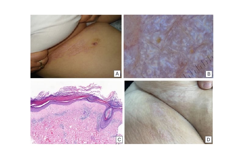
Figure 1: Dermatoscopic examination of the patient.
A) Clinical image of lichen sclerosus et atrophicus extending from the left inner thigh to the inguinal fold showing porcelain white crinkled violaceous patches. B) Dermatoscopic findings revealing brown follicular plugs, white lines, and a prominent rainbow pattern arranged over white polygonal cods on polarised mode (Dermlite DL4; DermLite, San Juan Capistrano, California, USA). C) Histologic findings revealing epidermal hyperkeratosis, atrophy, follicular plugs with basal vacuolar degeneration, and homogenous dense fibrosis in the papillary dermis with dense lymphocytic infiltrate beneath the fibrosis (haematoxylin and eosin stain, 7.3x magnification). D) Clinical image of lichen sclerosus et atrophicus showing regression of the lesions except mild atrophy and depigmentation after 2 years of topical steroid use.
DISCUSSION
Extragenital LSEA mostly occurs as multiple, oval, bluish–whitish macules or papules located on the neck, shoulders, and upper trunk. Lesions are predominantly asymptomatic. Itching is the most common complaint in symptomatic cases and leads to increased frequency of scratching followed by scarring. Diagnosis is mostly clinical; however, in ambiguous cases, dermatoscopy and histopathology lead to diagnosis. Dermatoscopic features of LSEA are characterised by white structureless areas, follicular plugs, purple dots, shiny white streaks, and peppering.1 In the authors’ patient, a prominent rainbow pattern arranged over white polygonal clods on polarised dermatoscopy was observed. The rainbow pattern has been reported in a few LSEA cases in the literature.2 In previous studies, this pattern has been associated with a rich vascular network; however, the authors’ histological examination did not reveal a vascular-rich pattern. The authors observed epidermal hyperkeratosis, atrophy, and homogeneous dense fibrosis in the papillary dermis and dense lymphocytic infiltrate below the fibrosis. Although the mechanism behind this appearance is not clear, it can be secondary to diffuse and dense homogenous fibrosis in the superficial dermis, giving a sharp and brighter appearance along with increased dermal capillaries and inflammatory cell infiltration.
CONCLUSION
Extragenital LSEA should be considered in the differential diagnosis in the presence of dermatoscopic rainbow pattern, shiny white lines, and follicular plaques along with clinical features.