ACCORDING to a recent study, the use of thermal imaging and the ALT-70 (asymmetry, leukocytosis, tachycardia, age ≥70 years) prediction model effectively distinguished between cellulitis and pseudocellulitis, thus limiting the frequency of misdiagnoses.
Cellulitis is a frequently misdiagnosed condition, with up to 30% of cases misdiagnosed due to the prevalence of largely indistinguishable maladies, termed pseudocellulitis. The threat to public health is not so much found in the misdiagnoses themselves, but in the prescription, and subsequent overuse, of antibiotics within the population.
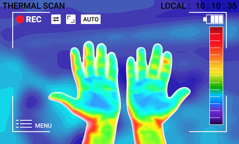
The study, a prospective diagnostic validation study, comprised 204 patients (mean age: 56.6 years; 121 male), 45.1% of whom had a consensus diagnosis of cellulitis, determined between 11th October 2018–11th March 2020. The maximum and mean temperatures of afflicted, and corresponding unafflicted, limbs were recorded using a thermal camera attached to an iPad (Apple, Cupertino, California, USA). ALT-70 scores indicate the presence or absence of cellulitis, with a score ≥5 indicating cellulitis requiring treatment, a score between 3–4 requiring consultation of a dermatologist, and a score ≤2 unlikely to be cellulitis.
Of the cohort, 12% had a score ≤2, 47% a score between 3–4, and 40% a score ≥5. Of those with a score ≥5, 40% were identified as having a false-positive ALT-70 result, indicative of pseudocellulitis. Between the two conditions, there were statistically significant discrepancies in all skin surface temperatures (mean temperature, maximum temperature, and gradients). The maximum temperature of the affected skin surface in cellulitis was recorded at 33.2 °C, versus 31.2 °C in pseudocellulitis (95% confidence interval: 1.3–2.7 °C; P<0.001). The sensitivity of ALT-70 and thermal imaging exceeded 90% (98.8% and 93.5%, respectively), where specificity was highly variable (22.0% for ALT-70; 95% confidence interval: 15.8–28.1%). Combining both methods showed the greatest specificity, at 53.9%.
Regarding their findings, the authors wrote: “Cellulitis results in skin surface temperature elevations significantly greater than in pseudocellulitis. Surface thermal imaging may represent a useful adjunct to the clinical assessment of cellulitis (reducing overdiagnosis), but additional validation of diagnostic performance and cutoff values in more diverse populations is needed.”
Reference
Pulia MS et al. Validation of thermal imaging and the ALT-70 prediction model to differentiate cellulitis from pseudocellulitis. JAMA Dermatol. 2024;DOI: 10.1001/jamadermatol.2024.0091.


