Speakers: Patrick Trevidic,¹ Lee Walker,² Benji Dhillon,3 Ayad Harb,⁴ Kieren Bong,⁵ Raul Cetto,⁶ Rémi Foissac,⁷ Sabrina Shah-Desai⁸
1. Expert 2 Expert, Paris, France
2. BCity Clinics, Liverpool, UK
3. Define Clinic, Beaconsfield, UK
4. Dr Ayad Harb Clinic, Bicester, UK
5. Essence Medical, Glasgow, UK
6. Clinic 1.6, London, UK
7. Polyclinique Saint George, Nice, France
8. Perfect Eyes Ltd., London, UK
Disclosure: All speakers have served as consultants for Teoxane.
Acknowledgements: Medical Writing assistance was provided by Pauline Maffert, Clinical & Medical Affairs Department, Teoxane, Geneva, Switzerland.
Support: The symposium and publication of this article were funded by Teoxane.
Citation: EMJ Dermatol. 2021;9[1]:42-50.
Meeting Summary
Recent years have witnessed significant advances in the understanding of facial anatomy. Extensive studies have uncovered the arrangement of soft-tissue layers underlying the skin and their mobility during expression.1,2 Meanwhile, increasing awareness of the facial vascular network has laid the foundation for lower risk cosmetic procedures.3-5 In this context, facial aesthetic surgery tends to lose ground to less invasive procedures that, in the right hands, may provide similar benefits through less traumatic, relatively painless treatments and minimal social downtime. Hyaluronic acid (HA) fillers have thus gained popularity and evolved from their ’wrinkle filling’ role to expand to facial contouring, volume augmentation, and profiloplasty.6-8 Despite their well-established efficacy and safety profile, achieving natural-looking results in these indications requires specific products and appropriate techniques adapted to patient age, gender, ethnicity, and facial dynamism.9-12 The ‘anatomy, techniques, products’ approach, which stems from these core principles, was presented during two symposia at the Aesthetic and Anti-aging Medicine World Congress (AMWC) 2021, focusing on HA filler treatments in the mid- and lower face.Symposium 1: Midface
Sabrina Shah-Desai, Patrick Trevidic, Lee Walker, Benji Dhillon, Ayad Harb, and Kieren Bong
Anatomy
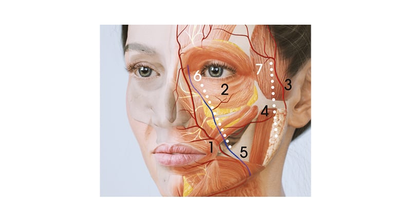
During the midface symposium, moderated by Sabrina Shah-Desai, Patrick Trevidic emphasised the necessity to understand the 3D vascular network of the face, considering the poor reliability of pre-injection aspiration.13 He proposed to delimit the injection area lateral to the mid-cheek groove and above the zygomatic line to respectively rule out risks of encountering the facial artery (FA), facial vein, and the transverse FA. As all branches of the infraorbital artery run down and medially in the face after emerging from the infraorbital foramen, staying lateral to the mid-cheek groove should also minimise risk of injury. Furthermore, small capillaries rising from the zygomatico FA and running up superficially are theoretically too small to constitute a potential HA embolus’ door (Figure 1).14-18

Figure 1: Vascular dangers of the midface and proposed safety lines delineating a relatively avascular area.
1) Facial artery; 2) infraorbital foramen; 3) superficial temporal artery; 4) transverse facial artery; 5) facial vein; 6) mid-cheek safety line; and 7) lateral safety line.
After having detailed the five-layered arrangement of the midface, including the skin, superficial fat, superficial musculoaponeurotic system, deep fat, and bone, a fourth dimension was introduced: facial dynamism.19 Showing the animation of a “smiling cadaver,” Trevidic illustrated the behaviour of deep versus superficial fat compartments upon muscle contraction. While the former sticked to the bone and remained static during movement, the latter moved along with the underlying muscle and soft tissue envelope.20
Of note, as augmenting the midface may also impact the perception of other areas of the face, patient assessment should include an evaluation of adjacent anatomical zones, including the temple, tear trough, and nasolabial fold.21
Lee Walker described how ageing causes an overall descent of the soft tissue envelope and how it affects each layer of the midface. In the dermis, the collagenous extracellular matrix is altered and elastin fibres disorganise as a result of intrinsic and extrinsic ageing.22 Meanwhile, superficial fat compartments undergo selective hypertrophy and lengthening; the underlying superficial musculoaponeurotic system becomes lax; facial muscles lengthen, thereby reducing the amplitude of movement; and all deep fat pads lose volume.22 Finally, bony remodelling in the face causes the glabellar, pyriform, and maxillary angles to decrease, allowing overlying soft tissues to rotate in an anterior direction.23 Seeing the face as a landscape, these combined changes culminate in the appearance of hypertrophic hills side-by-side with atrophic valleys.24 The most dramatic volume changes occur between 30 years and 60 years, with patients losing approximately 11% of their superficial fat and 18% of their deep fat.25
To avoid the pitfall of the overfilled midface and the appearance of ‘apple cheeks’ upon smiling, Walker explained the role of the transverse facial septum, which displaces upon smiling, thereby causing an anterior projection of deep midfacial fat compartments.26 Being aware of this mechanism, injectors are advised to ask their patient to smile repeatedly during the procedure.
Techniques and Products Tailored to Each Patient
To fully address the ageing process affecting all layers of the midface, a multilayering approach was recommended, aiming to compensate for deep and/or superficial volume loss depending on patient assessment, using appropriate techniques and products. Limiting the injections to the deep layer would require higher volumes to get the same lifting effect,27 while injecting high volumes or inadequate products superficially could lead to bumps and irregularities under the skin.28
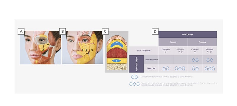
Products injected into each layer should be adequately chosen to mimic the original biomechanical properties and role of the fat, i.e., projection and support in the deep layer, and dynamic volumising in the superficial layer (Figure 2).

Figure 2: The multilayer anatomy of the midface spanning from bone to skin.
A) The static deep fat compartments: 1) mSOOF; 2) lSOOF; and 3) medial and lateral DMCF. B) The mobile superficial fat compartments: 1) IOF compartment; 2) MCF compartment; 3) MiCF compartment; and 4) NLF compartment. C) A multilayer treatment algorithm for filling 1) deep and 2) superficial fat layers of the mid-cheek with hyaluronic acid fillers adapted to specific patient needs, depending on age, gender, and skin type (D).
DMCF: deep medial cheek fat; IOF: infraorbital fat; lSOOF: lateral sub-orbicularis oculi fat; MCF: medial cheek fat; MiCF: middle cheek fat; mSOOF: medial sub-orbicularis oculi fat; NLF: nasolabial fat.
Furthermore, to ensure natural-looking outcomes, techniques and products should be fine-tuned to adapt to each patient anatomy. To illustrate this point, Benji Dhillon reviewed the ideals of facial beauty across ethnicity and gender, and anatomical differences influencing treatment plan.
While the male face is typically squarer than the female’s and less full in the anteromedial cheek, female attractiveness lies in the cheeks. It is commonly associated with a heart to oval shaped face, with a bizygomatic distance greater than the bigonial distance (versus the equivalent in men), prominent upper facial features, and lateral cheeks smoothly running into full medial cheeks.29
Ethnicity brings further complexity to these general notions. White people present narrower faces, with greater vertical height and projection in the upper face and midface, whilst Southeast Asian people have a wider face with shorter vertical height and are flatter in the medial maxilla.30,31 African-Caribbean people have a greater adiposity and skin thickness, a wider alar base, and ideally present an oval-shaped face.32 Finally, Middle Eastern women show well defined and full lateral cheeks, and an oval or square shaped face.33 However, some aesthetic ideals tend to evolve with globalisation, setting new aesthetic goals such as “ethnic optimisation,” “westernisation,” and “ethnic feature blending,” going against traditional beauty concepts.34
Live Demonstrations
Four expert injectors applied a tailored multilayering technique to the midface of patients from different ethnicities and ages, concretely showing how “each patient is unique.”
Focus on ethnicity: Middle Eastern and Southeast Asian patients
Middle Eastern female patient
Ayad Harb initiated live injections with a Middle Eastern female patient aged 32 years, concerned by the flatness of her midface giving her a tired look.
Objectively, the patient was perceived as young, without visible signs of deterioration of her volume or skin. She did present, however, with noticeable shadows and deficiencies in her midface and tear trough, and a lack of anterior projection. On the other hand, she had sufficient projection and contour in her lateral cheek. Therefore, the treatment aimed to enhance the anterior projection of her midface, fill her medial cheek, and further camouflage her tear trough, if still prominent after her midface treatment.
A 25 G cannula technique was used for injecting both the deep and superficial fat of the midface from a single-entry point on the zygoma.
Starting with the deep fat compartments, a strong projecting filler (TEOSYAL® Ultra Deep [HAUD]) was injected very slowly in small boluses in the medial sub-orbicularis oculi fat (mSOOF) and deep medial cheek fat (DMCF; 0.3 mL per compartment). The lateral SOOF (lSOOF) was left out in this patient already as she was showing good lateral projection.
In the superficial fat, another volumising but more dynamic filler (TEOSYAL RHA® 4 [RHA 4]) was selected to enhance the patient’s natural contour. Asking her to smile to see her cheeks animated helped to identify the areas to be filled. The infraorbital fat (prone to oedema and water retention) and the nasolabial fat (which hypertrophies with aging)35 were excluded from the treatment zone. The filler was placed evenly across her medial and middle superficial fat, injecting 0.3 mL per side.
Southeast Asian female patient
Dhillon performed the second live injection on a 37-year-old female patient presenting a complex blend of ethnicities, being half European, a quarter Taiwanese, and a quarter Korean.
She presented a heart to oval shaped face with an adequate ratio between bizygomatic and bigonial distances. She did show some degree of volume loss in the midface and pyriform fossa, though very limited considering her age and ethnicity. Upon smiling, her cheek filled up beautifully showing she was full in her medial superficial fat. Her youth and good skin quality were suitable indications for using a strong product in her deep fat compartments. In her case, treatment goals were thus to enhance the projection of her anterior medial cheek, and to improve the fullness of her subcutaneous submalar area.
Small boluses (0.1–0.2 mL) of HAUD were placed deep onto the periosteum in the mSOOF and DMCF, accessing the area with a 25 G cannula introduced from the zygoma. In this young patient with limited volume loss, no superficial injections were needed in the medial cheek.
However, superficial injections were performed with a cannula using a slightly lower entry point, targeting the area just below the zygomatic arch to correct its slight hollowness with little threads of RHA 4 (0.7 mL per side).
Finally, moving to the boundaries of the midface area, a small bolus (0.1 mL) of HAUD was injected deep onto the periosteum in the pyriform fossa, giving a little more reflection to this area.
Focus on age
Younger female patient
Kieren Bong treated a 32-year-old female with another specific background, as she was a professional athlete with very low body fat. She showed a mild lengthening of the lid-cheek junction, a slight infraorbital hollowness, and lacked some anterior projection and fullness.
Deep injections of HAUD were performed using a cannula technique from an entry point on the extension of the tear trough, slightly higher than the mid-cheek groove, located using the length of the cannula (50 mm). A first bolus was placed in the upper mSOOF, aiming to shorten the vertical height of the lower lid. To provide mild frontal and slight lateral projection to her midface, two other boluses were injected in the mSOOF and lSOOF, paying attention to avoid the malar mound. An overall volume of 0.6 mL of HAUD per side was needed.
A small amount of TEOSYAL® PureSense REDENSITY 2 was then injected in her tear trough to soften her infraorbital hollow with this specifically designed filler.36,37 The product was placed deep onto the bone, slightly above the orbicularis retaining ligament (ORL), and then massaged into place.
Lastly, superficial injections of RHA 4 were done from a second entry point, approximately 1 cm lateral to the lateral canthus, injecting 0.5 mL of product in linear threads.
Older female patient
Midface live demonstrations ended with a 49-year-old patient with more visible signs of ageing, volume loss, and skin laxity. She was thin, with low body fat and a more skeletonised appearance.
The ORL and zygomatico-cutaneous ligament (ZCL) were easily marked on her face. The ORL (superiorly) and ZCL (inferiorly) house the superficial infraorbital fat, which was prominent in this thin patient. This area of poor lymphatic drainage should always be avoided during a filling procedure to avoid worsening the bulging appearance of the infraorbital fat. The lSOOF lying deep between the ORL and the ZCL, lateral to the orbital line, was excluded from the treatment plan as lateralising this patient’s midface could have further deepened her temporal fossa and preauricular area.
Aiming to create a positive vector on the patient’s face, deep injections were performed in the mSOOF with RHA 4 (0.2 mL) and in the DMCF with HAUD (0.1 mL) using a needle bolus technique. In this thin-skinned patient, RHA 4 was appropriate to provide anterior projection in the mSOOF, which efficiently translates filler volume into surface projection. In the DMCF, where the surface-volume coefficient is lower, the stiffer, more cohesive HAUD gel was preferred to get the desired projection with minimal volume.38
The second treatment step aimed to fill the medial and middle superficial fat with RHA 4. A 25 G cannula was advanced in the submalar area from a medial entry point. The cannula was considered safer as the superior emerging branch of the transverse FA lies in the subcutaneous plane, approximately a finger under the zygoma.5
Symposium 2: Lower Face
Patrick Trevidic, Raul Cetto, Benji Dhillon, and Rémi Foissac
Anatomy
The ‘anatomy, techniques, products’ approach applied to the lower face was illustrated during four live injections in indications covering the anterior and posterior parts of the lower third of the face.
Key vascular dangers of the lower face were summarised by Trevidic. He drew the path of the FA and facial vein, crossing the inferior border of the mandible anterior to the insertion of the masseter and deep to the platysma, with the main trunk of the FA going up to the nasolabial fold while its submandibular branch gives the mental ascending branch emerging in the chin, approximately 6 mm from the midline, within the submuscular plane (Figure 1).39-41
He also showed the three fixed points on the lower face where soft tissues are tightly attached to the bone: the mandibular ligament, parotidomasseteric ligament framing the jowl, and mandibular angle.42-44 Their presence may become evident during ageing with tissue ptosis, resulting in a disrupted jawline.
The arrangement of soft tissue layers is also quite different between the anterior and posterior lower face. The chin is the most dynamic part, where three paired muscles can be found on each side of the mouth: the depressor anguli oris, depressor labii inferioris, and mentalis. In this highly dynamic area, there is a traditional five-layered arrangement just like in the midface. By contrast, lateral to the mandibular ligament lies the static part of the lower face, i.e., the jawline and the gonial angle, where the masseter is directly attached to the bone without intermediate fat.2,45
Techniques and Products Tailored to Each Patient
Age and gender specificities
Desirable aesthetic features in the lower face include a projected chin and a defined line from the gonial angle to the chin, contributing to a youthful look.7 However, there are gender specificities to be considered before establishing a treatment plan. Males typically have a squarer face than females, with a sharper jawline and a more pronounced jaw angle, whereas females present a more oval facial shape. The chin is also larger in males, whereas in females it is rounder and more subtle.7
Younger female patient
In the first live demonstration, a 43-year-old female patient was treated with the aim of improving her chin projection and redefining her mandibular line.
Chin projection was obtained by placing 0.6 mL of HAUD in the midline of her chin, going deep, straight onto the periosteum using a needle, the bevel facing the bone.
Afterwards, RHA 4 was injected in the paramedian area using a fan-technique, introducing a cannula in the superficial plane from an entry point located at the bottom of the marionette line. Four to five linear threads were placed along the mandible to recreate the anterior jawline, and superiorly to improve the marionette line and corner of the mouth. The posterior part of the jawline was addressed using a superficial cannula technique from an entry point posterior to the jowl, advancing in direction of the mandibular angle. One syringe (1.2 mL) of RHA 4 was needed to improve one side of the face. All superficial injections were followed by post-injection moulding to smooth the product into place.
Younger male patient
Gender differences required an adaptation of product and volumes in Raul Cetto’s 38-year-old male patient.
Anatomical specificities were first regarded from an anterior perspective. In a male face, the width of the chin ideally corresponds to the width of the mouth instead of the intercanthal distance in females, and the bigonial distance should be close to the bizygomatic width. From a sagittal perspective, the chin tends to project further in males than in females. A squarer face is considered attractive, and angles are typically sharper. The mandibular line should be straight and uninterrupted, and often needs to be elongated to accentuate masculine traits in a male’s face. Asking the patient to slide his teeth forward helps to visualise how far it can be improved.7,45
The first treatment step aimed to improve chin projection. To respect the width of the male chin, HAUD was injected in two supraperiosteal boluses (0.2–0.3 mL each) on both sides of the midline, instead of one central bolus. The superficial layer of the chin was then filled with RHA 4 using a cannula entered at the position of the mandibular ligament, delivering half a syringe on each side of the chin.
The main goal of the jawline treatment was to increase the bigonial width by placing a deep bolus of HAUD (0.3 mL) against the bone at the mandibular angle. Finally, using a cannula advanced from the mandibular angle, superficial injections of RHA 4 were completed to elongate the posterior jawline.
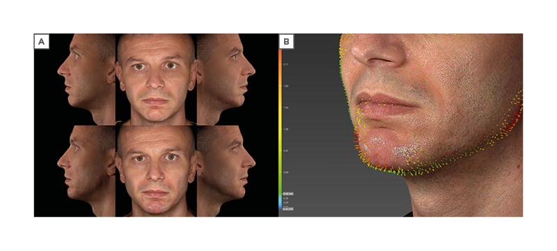
Before and after photographs can be seen in Figure 3.

Figure 3: Photographs of Cetto’s 38-year-old male patient taken with the VECTRA® H2 3D imaging system (Canfield Scientific, Parsippany, New Jersey, USA).
A) Before (upper) and after (lower) photographs of the patient. B) Visualisation of volume changes after treatment. The orange and red arrows indicate areas of volume augmentation.
Older female patient
Dhillon‘s 49 year-old patient had strong masseters, giving her a square face and a large bigonial width. This was considered in the treatment plan to avoid masculinising her. Her gonial angle seemed to match with female standards (120–130°), hence did not require much reconstruction.46 However, she presented mild jowling and lacked definition in her posterior jawline.
The jowl was demarcated as a ‘no go’ area, to concentrate treatment efforts on areas posterior and anterior to it. Entering from the jowl, a cannula was advanced in the subcutaneous plane in direction of the gonial angle to perform a fanning technique, depositing linear threads of RHA 4 while retracting the cannula above and below the mandibular border, to blend the mandibular line into the jowl. The same approach was used in the anterior part of the jawline, entering in the opposite direction. A total of 1.7 mL of RHA 4 per side provided the desired mandibular line and angle definition.
The chin was treated from a single-entry point in the midline. In the paramedian area, 0.6–0.7 mL of TEOSYAL RHA® 2 was fanned superficially in each side, opting for this spreadable and highly stretchable gel to limit risks of irregularities in this thin-skinned patient. To further enhance chin projection, a 0.2 mL bolus of RHA 4 was deposited supraperiosteally in the midline.
The whole treatment required an overall 4 mL of filler, resulting in a straighter jawline, greater definition, and light reflection.
Chin retrogenia
The last treatment session aimed to illustrate the treatment of congenital chin retrogenia in a 28-year-old female patient. Rémi Foissac used the Gonzales-Ulloa line to show the effect of chin retrusion on his patient’s profile, particularly on her nose, which seemed abnormally large.
To project her chin, a whole syringe HAUD was injected in deep boluses onto the periosteum of the midline and paramedian zones, using a cannula introduced from the midline. The subcutaneous plane was filled using a fanning technique, with a cannula entered from the lateral chin, delivering half a syringe of RHA 4 on each side of the chin.
Conclusion
The perception of a human face and, therefore, of a personality, is strongly impacted by the appearance of the midface and lower face. Age, gender, and ethnicity influence both the anatomy and the aesthetic ideals associated to these areas. HA fillers may be used to provide a more harmonious or youthful look but should be selected and injected with thorough consideration to each patient’s anatomy and background, keeping in mind the fourth dimension of the face, i.e., dynamic expression.


