Abstract
Introduction: Diabetic emergencies such as diabetic ketoacidosis (DKA) are life-threatening complications, often precipitated by infections or illnesses such as COVID-19.
Case presentation: A 55-year-old African American female presented to their primary care physician, complaining of fatigue, dehydration, decreased appetite, hypersomnia, and sudden weight loss, and a past medical history of Type 2 diabetes. They had a glucose level of >15 mmol/L and ketone level of >16 mmol/L; they were immediately sent to the emergency department for assessment of DKA. There, the patient tested positive for COVID-19. They had a glucose level of 361 mg/dL, a pH of 7.11, a bicarbonate level of 10 mEq/L, a sodium level of 125 mEq/L, a potassium level of 3.9 mEq/L, a chloride level of 95 mEq/L, an anion gap of 20, and a positive ketone level. Over the next few days, the patient’s condition got worse; their chest CT scan showed ground-glass opacities with consolidations in the middle and inferior lobes of the lungs bilaterally, along with interlobular septal thickening, which are consistent with an atypical infection, respiratory distress, and pneumonia. The patient was on intravenous fluids, insulin therapy and empirical antibiotics for the next few weeks, and eventually recovered.
Discussion: Factors precipitating DKA in patients with diabetes in the setting of COVID-19 are: the increased secretions of stress hormones that counter the effects of insulin and increase blood glucose levels, and the ways in which severe acute respiratory syndrome coronavirus 2 interacts with human cells, leading to pancreatic islet cell damage.
Conclusion: Diabetes and COVID-19 intensify each other’s complications in patients diagnosed with both.
INTRODUCTION
The severe acute respiratory syndrome coronavirus 2 (SARS-CoV-2) outbreak began in China in December 2019, and the World Health Organization (WHO) declared COVID-19 as a pandemic in March 2020.1 Since then, there have been 146,054,107 positive cases and 3,092,410 deaths globally to date in April 2021.2 Recent COVID-19 studies have shown that individuals above the age of 60, or with underlying health conditions such as obesity, diabetes, chronic kidney disease, cardiovascular disease, and cancer, are at a higher risk of severe illness and mortality.3 A study conducted in New York, USA, showed that the prevalence of diabetes was higher in patients with COVID-19 who were hospitalised (34.7%) versus recovering from home (9.7%).4 Another study based in England, UK, revealed that among patients with COVID-19 who have passed away at hospital, 32% had Type 2 diabetes and a 2.03 times higher rate of mortality, and 1.5% had Type 1 diabetes and a 3.5 times higher rate of mortality.5 Another study conducted in China showed that the prevalence of diabetes amongst the general population was 8.2%, and increased to 34.6% in patients with COVID-19.6 Diabetic emergencies like diabetic ketoacidosis (DKA) are life-threatening complications, often precipitated by infections or illnesses.7 Currently, there is limited data on the correlation between COVID-19 and DKA. The authors report a case of a 55-year-old African American female with COVID-19, who developed DKA.
CASE PRESENTATION
A 55-year-old African American female presented to her primary care physician (PCP), complaining of fatigue, dehydration, decreased appetite, hypersomnia, and sudden weight loss. They denied any fever, cough, sore throat, shortness of breath, headache, runny nose, vomiting, diarrhoea, and loss of taste or smell. Their past medical history included schizophrenia, Type 2 diabetes, hyperlipidaemia, and vitamin D and zinc deficiency. They were taking risperidone 25 mg intramuscular Q14, metformin 1,000 mg orally twice daily (PO BID), empagliflozin 25 mg once each morning (PO QAM), insulin glargine 25 units by single-carrier quadrature amplitude modulation rosuvastatin 40 mg orally at bedtime, vitamin D 2,000 units PO QAM, zinc gluconate 50 mg once daily (PO QD), and acetaminophen 325 mg every 4 hours, as needed.
Examination by the PCP showed that the patient’s vitals were stable. They had a blood pressure of 132/82 mmHg, a heart rate (HR) of 82 bpm, a respiratory rate of 16 breaths/min, and an oxygen saturation of 98%. The urine dipstick test showed a glucose level of >15 mmol/L and ketone level of >16 mmol/L. The patient was immediately sent to the emergency department (ED) for assessment of DKA. Upon arrival at the ED, the patient’s vitals remained stable. They had a blood pressure of 138/82 mmHg, an HR of 80 bpm, a respiratory rate of 16 breaths/min, and an oxygen saturation of 98%. A reverse transcription-PCR test was conducted as part of routine COVID-19 testing, and the patient tested positive for COVID-19. They were immediately moved into isolation, where further work-up was completed. Blood work and urinalysis were consistent with DKA; the former showed a glucose level of 361 mg/dL, a pH of 7.11, a bicarbonate level of 10 mEq/L, a sodium level of 125 mEq/L, a potassium level of 3.9 mEq/L, a chloride level of 95 mEq/L, an anion gap of 20, and the latter was positive for ketones. The patient was started on intravenous (IV) fluids, insulin therapy, and empirical antibiotics for the management of DKA and COVID-19. They were then transferred to the intensive care unit (ICU) for further care.
Over the next 3 days, the patient’s condition worsened. The ECG showed sinus tachycardia, probable left atrial enlargement, and borderline T-wave abnormalities; the patient was persistently tachycardic with HRs ranging from 100–140 bpm. The CT scan without IV contrast showed ground-glass opacities with consolidations in the middle and inferior lobes of the lungs bilaterally, along with interlobular septal thickening, which are consistent with an atypical infection, respiratory distress, and pneumonia. The peripheral doppler ultrasound ruled out deep vein thrombosis and the CT pulmonary angiogram ruled out pulmonary embolism. The patient had nasogastric and endotracheal intubation. They continued on IV fluids, insulin therapy, and empirical antibiotics.
Eventually, the patient’s DKA and COVID-19 resolved with standard management, and they were finally discharged home 1 month later. Their discharge prescription included risperidone 25 mg intramuscular Q14, metformin 1,000 mg PO BID, linagliptin 5 mg PO QAM (new medication), insulin glargine 25 units by single-carrier quadrature amplitude modulation, rosuvastatin 20 mg orally each bedtime (lowered dosage), ezetimibe 10 mg PO QAM (new medication), bisoprolol 5 mg PO QAM (new medication), vitamin D 2,000 units PO QAM, zinc gluconate 50 mg PO QD, multivitamin with iron 1 tablet PO BID (new medication), and acetylsalicylic acid 81 mg PO QD (new medication). The patient was referred to a diabetes educator and an endocrinologist for further follow-up.
The day after discharge from the ICU, the patient visited their PCP, who reviewed their ED and ICU notes, discharge prescription, and referrals. The patient was feeling better and was not in any distress. The PCP noted that the patient had a case of DKA precipitated by COVID-19.
DISCUSSION
It has been established that patients with diabetes are at increased risk of COVID-19 infection, ICU admission, and mortality.3-6 Multiple explanations have been postulated for this predisposition. Dysregulated immune responses in patients with diabetes lead to a higher susceptibility to infectious diseases.8 Furthermore, in vitro and animal studies show that hyperglycaemia facilitates local viral replication in the lungs and impairs antiviral immune responses.9,10 Conversely, fasting blood glucose levels are higher in patients with COVID-19. Additionally, higher levels of IL-6 seen in diabetes predispose these patients to a greater risk of a cytokine storm, which may lead to critical illness.8 Thus, diabetes and COVID-19 intensify each other’s complications in patients diagnosed with both.
The patient presented in this case was diagnosed with DKA that was likely precipitated by COVID-19. Although the patient did not present with typical COVID-19 symptoms, their reverse transcription-PCR test results prove the diagnosis. In addition, their chest CT scan results are consistent with typical COVID-19 pneumonia appearance, with a sensitivity of 73.5% and specificity of 82.8%, as per the Radiological Society of North America (RSNA).11 A study from China highlighted 58 COVID-19 cases characterised by positive chest CT scanning in which the patients were asymptomatic.12 Furthermore, the absence of other possible triggers for the development of DKA is suggestive that it was likely provoked by COVID-19.
DKA is a lethal complication of diabetes characterised by anion gap metabolic acidosis and the accumulation of ketone bodies due to uncontrolled blood glucose levels. This typically occurs in Type 1 diabetes but has also been seen in Type 2 diabetes, especially in the presence of an infection. The resistance or deficiency of insulin increases blood glucose levels to a range of 350 mg/dL (19.4 mmol/L) to 450 mg/dL (27.8 mmol/L), and up to 800 mg/dL (44 mmol/L).13 Clinically, the patient will present with decreased alertness, nausea, vomiting, dehydration, Kussmaul breathing, and abdominal pain. The patient’s blood glucose level on admission to the ED was 361 mg/dL, which was consistent with DKA. Furthermore, blood work showed a low pH, low bicarbonate level, and an elevated anion gap metabolic acidosis. The inability to use glucose due to insulin resistance and deficiency leads to enhanced lipolysis, and the primary source of energy shifts from glucose breakdown to fatty acid breakdown. Ultimately, there is an abundance of acetyl coenzyme A, which is converted to acetoacetic acid, and further reduced to β-hydroxybutyric acid. Both of these ketone acids contribute to anion gap metabolic acidosis and can be detected on urinalysis, as seen in this patient.
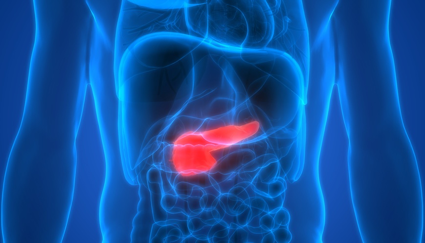
Since the beginning of the COVID-19 pandemic to date, some cases of DKA precipitated by COVID-19 have been reported. Multiple explanations to this pathophysiology have been proposed. One of the most common precipitating factors of DKA is infections or illnesses.14 They cause increased secretions of stress hormones such as catecholamines and cortisol, which counter the effects of insulin and increase blood glucose levels.15 Some studies have shown that patients with COVID-19 have significantly higher levels of cortisol, which may induce DKA if they are also diabetic.16 Another factor precipitating DKA in patients with diabetes may be the ways in which SARS-CoV-2 interacts with human cells. The spike protein receptor binding domain on SARS-CoV-2 binds to the angiotensin converting enzyme 2 receptors in humans; the host cell then internalises the receptor along with the bound virion, allowing it to become effective and cause cellular damage.17,18 Angiotensin converting enzyme 2 receptors are found on multiple organs throughout the body, including the pancreatic islet cells; as a result, SARS-CoV-2 binds to these pancreatic islet cells, leading to insulinopenia and increased risk for DKA.19
CONCLUSION
The 55-year-old patient with a medical history of Type 2 diabetes developed DKA in the setting of COVID-19. Patients with diabetes are known to have an increased severity of COVID-19 infection and COVID-19 infection is known to precipitate DKA. Therefore, diabetes and COVID-19 intensify each other’s complications in patients diagnosed with both.


