Abstract
Spontaneous carotid artery dissection in young patients can be caused by vascular structural abnormalities secondary to connective tissue disorders, or minor or no trauma. This is a rare case of a 37-year-old woman who had carotid artery dissection from the minor trauma of vomiting, and also had anterior and posterior cerebral infarcts via embolisation from the carotid artery dissection. She presented with multiple generalised and focal neurological symptoms; therefore, various neurological disorders were considered, which were then excluded. She was subsequently diagnosed with ischaemic stroke, which led to the diagnosis of carotid artery dissection, and managed with antiplatelet therapy. A further detailed history revealed an underlying diagnosis of Ehlers-Danlos syndrome. Consideration of spontaneous carotid artery dissection and ischaemic stroke or transient ischaemic attack in patients presenting with neurological symptoms, with a past medical history or family history of a connective tissue disorder or other condition predisposing vascular fragility, can lead to timely diagnosis and treatment. Young patients with spontaneous carotid artery dissection, and no significant medical or family history or trauma to indicate a cause, should be screened for connective tissue disorders and considered for genetic testing.
Key Points
1. Spontaneous carotid artery dissection is a very rare complication of hypermobile Ehlers-Danlos syndrome, caused by vascular structural abnormalities secondary to connective tissue disorders or by minor trauma such as vomiting.2. Young patients with spontaneous carotid artery dissection and no vascular risk factors should be screened for connective tissue disorders and considered for genetic testing. A detailed past medical history and family history should also be obtained to try and identify an underlying cause.
3. Carotid artery dissection is a recognised cause of anterior circulation ischaemic stroke or transient ischaemic attack. It can also, rarely, cause posterior circulation infarction or ischaemia due to an anatomical variant of a fetal posterior cerebral artery, or by embolisation from the dissection via a dominant posterior communicating artery.
INTRODUCTION
Carotid artery dissection (CAD) can arise from traumatic, spontaneous, and iatrogenic causes. Spontaneous CAD is generally rare, but a relatively common cause of ischaemic stroke in people under the age of 50 years.1 Certain connective tissue disorders (Ehlers-Danlos syndrome [EDS], Marfan syndrome, fibromuscular dysplasia, and osteogenesis imperfecta), and infectious and inflammatory disease, all increase the risk of spontaneous CAD due to associated vascular structural abnormalities.1 Spontaneous CAD can also be caused by minor trauma.
EDS is a rare, genetic, and heterogenous group of connective tissue disorders characterised by tissue fragility and hypermobility.2 Vascular EDS is commonly associated with significant arterial wall weakness, therefore predisposing individuals to CAD.1,3 However, spontaneous CAD is a rare complication of the other subtypes, such as hypermobile EDS.4 Due to vessel fragility in patients with EDS, dissections are liable to occur spontaneously or after minor trauma.1,5
CAD is a recognised cause of anterior circulation infarction. However, in rare cases, CAD can lead to posterior circulation infarction.
This case report illustrates multiple rare medical occurrences in one individual. Spontaneous CAD occurred in a woman with hypermobile EDS, and caused anterior and posterior circulation infarction. It highlights the importance of obtaining a detailed history of symptoms, particularly past medical history and family history, to make a timely and accurate diagnosis. Additionally, it prompts consideration of screening for connective tissue disorders and genetic testing for young patients with no significant medical history who present with spontaneous arterial dissection.
CASE PRESENTATION
A 37-year-old woman presented to the emergency department with a 24-hour history of vomiting, abdominal pain, and myalgia after travelling abroad. She was diagnosed with gastroenteritis and discharged. She presented again 3 days later with confusion, agitation, headache, visual hallucinations, right visual field loss, photosensitivity, and transient right-sided weakness with persistent altered sensation. Her past medical history included depression and irritable bowel syndrome. She smoked marijuana, but no tobacco, and was teetotal. A clinical examination was unremarkable.
Diagnosis
On initial presentation to the emergency department, the patient was diagnosed with gastroenteritis, having just returned from the Caribbean. Three days later, she presented with multiple neurological symptoms, non-specific and focal. Inflammatory markers were mildly raised (white cell count: 12.3×10⁹ /L; C-reactive protein: 42 mg/L). A CT brain scan was normal. Differential diagnoses included migraine with aura, infectious or autoimmune encephalitis, multiple sclerosis, and functional neurological disorder. Based on her symptoms, recent travel, and raised inflammatory markers, she was treated for infectious encephalitis.
A lumbar puncture was performed. Cerebrospinal fluid (CSF) analysis revealed a white cell count of 3 cells/mm3, a glucose level of 3.1 mmol/L (no serum glucose for comparison), and a protein level of 263 mg/L, which were normal. Bacterial culture, virology PCR, and Cryptococcus antigen were all negative, excluding infectious encephalitis. Oligoclonal bands and N-methyl-d-aspartate (NMDA) receptor antibodies were also negative, excluding multiple sclerosis and autoimmune encephalitis, respectively. The CSF result was therefore entirely normal.
MRI of the brain was subsequently carried out, which showed acute ischaemia in the left hippocampus and left high parietal and occipital regions (Figure 1A). CT angiography showed a left cervical CAD with significant stenosis and a small dissecting aneurysm (Figure 1B). There was no vertebral artery dissection. The patient had no preceding trauma. Her only vascular risk factors were smoking marijuana and her grandmother having a stroke in older age.
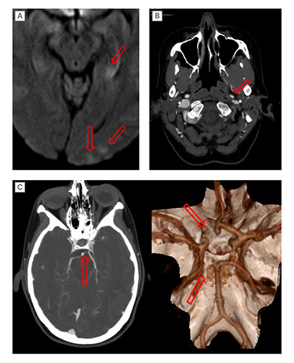
Figure 1: Imaging demonstrating cerebral ischaemia, arterial dissection, and dominant PComA.
A) MRI diffusion-weighted restriction image demonstrating faint diffusion restriction in the left hippocampal body/tail and left occipital cortex. B) CT angiogram axial imaging demonstrating a non-occlusive dissection of the left cervical carotid artery with a small, distal dissecting aneurysm. C) CT angiogram MIP demonstrating a small left P1 posterior cerebral artery segment as a result of a dominant PComA, and surface rendered model of the circle of Willis (viewed from above and posterior) demonstrating congenitally absent left A1 and P1 segments with the left internal carotid artery supplying the left posterior cerebral artery.
MIP: maximum intensity projection; PComA: posterior communicating artery.
Initial Management Plan and Outcome
The patient was admitted following her second presentation to the emergency department with neurological symptoms. She was commenced on intravenous antimicrobial therapy (ceftriaxone and aciclovir) for suspected infectious encephalitis. Once this diagnosis was excluded, further investigations were performed, which led to the diagnosis of ischaemic stroke and CAD. The patient was started on high-dose aspirin (300 mg once daily) for 14 days and then clopidogrel 75 mg once daily thereafter. She was referred to the stroke team. The patient was discharged 7 days after admission.
Case History
During assessment by a stroke specialist, a history of EDS was identified. The patient was diagnosed with Type III (hypermobile) EDS aged 18 years after having recurrent knee subluxations and dislocations without trauma from the age of 13 years, as well as skin hyperextensibility. Her mother also had EDS, with unspecified cardiac complications, so she was not offered genetic testing but had a skin biopsy. Management of her EDS was conservative, which included supportive exercises and swimming. Surgery was not recommended due to the increased risk of developing osteoarthritis at a young age. As she had skin hyperextensibility, she had surgery for the removal of excess skin after significant weight gain followed by weight loss as an adult.
The underlying connective disorder with minor trauma from vomiting was thought to cause the CAD. Posterior circulation ischaemia was an unexpected consequence of embolisation from the CAD via a dominant posterior communicating artery (PComA; Figure 1C). An interval CT angiogram 5 months later showed near complete remodelling of the previously dissected left cervical internal carotid artery. The dissecting aneurysm had resolved with now normal arterial calibre. Although the cervical internal carotid artery was normal on a CT angiogram at 9 months, given the EDS rendering vascular susceptibility and risk of further arterial dissection, clopidogrel was continued.
DISCUSSION
CAD is rare and contributes to 2% of ischaemic strokes overall, but is a common cause of ischaemic stroke in young people under the age of 50 years, accounting for 20–25% of cases.3,5 Spontaneous CAD is even less common, and can occur as a result of minor trauma, such as neck manipulation, coughing, sneezing, and vomiting.1,5 Certain connective tissue disorders, as well as infectious and inflammatory diseases, can cause spontaneous CAD by compromising the structural integrity of arterial walls.1 The combination of vascular fragility secondary to EDS and minor trauma from vomiting were the reasons for CAD leading to strokes in this case.
Patients with CAD often present with non-specific symptoms, such as headache, neck pain, and dizziness.2 More typical symptoms of CAD are ipsilateral cervical pain (with headache), ipsilateral Horner’s syndrome, transient ischaemic attack (TIA), or ischaemic stroke.5 In this case, the patient presented with multiple generalised and focal neurological symptoms. Furthermore, she had no major vascular risk factors, and the past medical history of EDS was not identified during her initial assessment. Various neurological diagnoses were considered, but not CAD or ischaemic stroke. Based on the patient’s symptoms and initial investigation results, encephalitis was thought to be the more likely diagnosis, which is why the lumbar puncture was performed before a brain MRI. If the history of EDS was elicited on admission, this may have prompted consideration of the patient’s typical and focal symptoms, which could have led to earlier diagnosis of CAD and ischaemic stroke, and an invasive lumbar puncture could have been avoided.
The gold standard investigation for diagnosing CAD is digital subtraction angiography; however, CT angiography is primarily used due to its high specificity and sensitivity, availability, speed, and because it is non-invasive.1,3 In this case, as soon as the diagnosis of ischaemic stroke was made from the brain MRI, CT angiography was carried out, which revealed CAD.
Antithrombotic treatment with either antiplatelet or anticoagulant medication remains the mainstay of treatment for CAD.3 The duration of treatment varies between individuals. It is usually given for 3–6 months, but may be longer depending on vascular risk factors and the extent of remodelling of the dissection.3 For patients with CAD with recurrent TIAs or ischaemic stroke despite medical therapy, haemodynamic cerebral hypoperfusion, or expanding pseudoaneurysm (associated with CAD), endovascular treatment should be considered, such as carotid stenting or coil embolisation of a pseudoaneurysm.1,3
EDS is a rare, genetic, and heterogenous group of connective tissue disorders characterised by tissue fragility and joint hypermobility, of which there are 13 subtypes.2 Specific subtypes, notably vascular EDS (previously known as Type IV EDS), are commonly associated with significant arterial wall weakness, which predisposes individuals to arterial dissection spontaneously or after minor trauma.1,2 Spontaneous CAD is a frequent complication of vascular EDS,2,6 and so this EDS subtype is more commonly associated with ischaemic stroke or TIA. Young patients with spontaneous CAD and ischaemic stroke or TIA with few or no vascular risk factors should be screened for connective tissue disorders and considered for genetic testing.3
Vascular EDS can cause other arterial complications, which include rupture of medium and large-sized arteries, aneurysm formation, and arteriovenous fistulas.2,6 It is also associated with non-vascular complications, such as hiatus hernia, organ perforation (gastrointestinal), and rectal and uterine prolapse.7 Most patients with vascular EDS tend to develop arterial complications by the age of 40 years.2 This case highlights that the occurrence of arterial complications at a young age is also applicable to other EDS subtypes, such as hypermobile EDS.
Vascular EDS is diagnosed by genetic testing, where mutations in the COL3A1 gene yield abnormal Type III collagen protein.2,6 As Type III collagen is also found in the skin and joints, patients with vascular EDS present with thin, translucent skin, easy bruising, and hypermobility of small joints.2 If the patient in this case did not have an established diagnosis of EDS, it would be prudent to carry out a further assessment and genetic testing for connective tissue disorders to identify the cause of spontaneous CAD. By making a diagnosis of a connective tissue disorder, appropriate management and surveillance can then be initiated.7
Hypermobile EDS is the most common subtype of EDS, which was diagnosed in the patient at the age of 18 years. Although it is an inherited disorder, it is the only subtype of EDS that cannot be diagnosed via genetic testing, as no genetic mutations have been identified.8 It predominantly occurs in females. The diagnosis of hypermobile EDS relies on clinical examination for evidence of a systemic connective tissue disorder; identifying generalised joint hypermobility of specific joints, such as knees and elbows, using the Beighton scale; and a family history of hypermobility and EDS.4,8 In this case, the patient’s history of recurrent knee subluxations and dislocations and family history led to her diagnosis. She had a skin biopsy, which can show ultrastructural skin connective tissue abnormalities,9 and this method can be used to support the diagnosis of hypermobile EDS.
In this case, the patient inherited EDS from her mother, who also had associated cardiac complications, which were unspecified. Hypermobile EDS can be associated with aortic root dilatation and mitral valve prolapse.4,8 Whilst hypermobile EDS is frequently characterised by joint hypermobility and skin hyperextensibility, patients with hypermobile EDS may, rarely, have extra-aortic complications, with cases of spontaneous coronary artery dissection and CAD being reported,4 as evidenced by this case. The patient had recurrent joint hypermobility issues during adolescence, as well as a positive family history, which led to her diagnosis of EDS. She had no prior history of vascular or other extra-arterial complications until the spontaneous CAD at the age of 37 years.
There is no definitive treatment for vascular EDS. Management is centred on symptomatic and preventative treatment, and genetic counselling.2 Preventative treatment can include lifestyle modification, such as avoiding certain physical activities to minimise injury and therefore reduce risk of arterial dissection and organ rupture,7 and control of vascular risk factors, especially hypertension.2,7 Patients with vascular EDS who have an ischaemic stroke or TIA are usually treated with antithrombotic therapy, either antiplatelet or anticoagulant, for 3–6 months.2 Antithrombotic therapy can be stopped if there is complete resolution of the CAD, but it can be continued to prevent new or recurrent ischaemic stroke.2 In this case, clopidogrel was continued because of concerns about the high risk of recurrence of ischaemic stroke or TIA due to the underlying EDS, despite the hypermobile subtype, and spontaneous CAD. Endovascular treatment or surgery for CAD and any other arterial dissection in patients with vascular EDS is not recommended due to the high risk of complications from existing vascular fragility, such as dissection or ischaemic stroke,2,10 unless the event is life-threatening. Stenting is a treatment option for recurrent CAD and recurrent ischaemic stroke or TIA, but this would require careful consideration.
CAD is a recognised cause of anterior circulation infarction, usually affecting the middle and anterior cerebral artery territories, while vertebral artery dissection commonly causes posterior circulation infarction. However, there are rare occurrences of posterior circulation infarction secondary to CAD. This can be due to underlying anatomical variants. This includes fetal origin of the posterior cerebral artery (PCA), whereby the PCA originates directly from the internal carotid artery, so that the posterior circulation is provided by the PComA.11 However, there are cases where PCA territory infarcts can be caused by embolisation from a CAD via a patent or dominant PComA.11
The main limitation of this case is that the diagnosis of EDS was made several years ago and before the wider availability of electronic medical records. Until the patient had CAD and ischaemic stroke, the EDS did not cause her significant problems in adulthood, only during adolescence. Therefore, the history of diagnosis and management of EDS was solely obtained from the patient’s recall of events 20 years ago, as her medical records from that period could not be accessed for specific information about clinical assessments, investigations, and management.
CONCLUSION
There were multiple rare medical occurrences in one individual resulting in an extremely rare case. Spontaneous CAD is rare, but commonly causes ischaemic stroke in patients under the age of 50 years. CAD can have non-specific presentations, which can lead to delayed diagnosis. A detailed history, particularly past medical history and family history, is vital for guiding appropriate investigations in making a correct diagnosis. Young patients diagnosed with ischaemic stroke or TIA with few or no vascular risk factors should have CT angiography for possible arterial dissection. Connective tissue disorders, such as EDS, are rare and can cause vascular structural abnormalities that can result in spontaneous CAD, especially with no or minor trauma, as demonstrated in this case. Young patients with spontaneous CAD and no relevant medical history or vascular risk factors should be screened for connective tissue disorders and considered for genetic testing. CAD has the potential to cause posterior circulation infarction in rare circumstances, either by embolisation from the dissection via a dominant PComA or an underlying vascular anatomical variant.