Abstract
Background: Marfan syndrome (MFS) and autosomal dominant kidney disease (ADPKD) are two separate genetic disorders. The author describes the case of a young male with ADPKD who incidentally had Marfan-like features. A literature review was carried out to see if these two disorders could be linked.
Case presentation: A young male presented for incidentally found renal cysts. Kidney function was well preserved, but the patient had positive family history of ADPKD. During routine follow-up, a history of aortic valve disease was mentioned. This, along with the patient’s tall, lean stature and long extremities raised the concern for MFS. A detailed physical examination and workup by other specialists confirmed a clinical diagnosis of MFS. They had no known family history of MFS. The patient has been followed at Associates in Kidney Care, Des Moines, Iowa, USA, for the past 2 years.
Discussion: There are several reports of overlap of ADPKD and connective tissue disorders with an overlap of vascular disorders. ADPKD and MFS are caused by totally different mutations. However, the literature review showed that vascular abnormalities and connective tissue diseases may be more common with ADPKD. Studies have shown that there could be a common signalling pathway for connective tissue disorders when both genes are affected simultaneously. Further research is needed to identify these pathways. More frequent screening of vascular abnormalities might be warranted in those with both phenotypes.
Key Points
1. The co-occurrence of Marfan syndrome (MFS) and autosomal dominant polycystic kidney disease (ADPKD) is uncommon, as these disorders affect 1 in 3,000–5,000 and 1 in 500–5,000, respectively.2. MFS and ADPKD result in systemic connective tissue symptoms, which can appear to overlap; however, more research is needed to identify genetic pathways that could cause this overlap in these disorders.
3. According to the author, patients with ADPKD and aortic vascular abnormalities should also be evaluated for the co-occurrence of other connective tissue diseases such as MFS.
BACKGROUND
Marfan syndrome (MFS) is an autosomal dominant connective tissue disorder with an incidence of 1 in 3,000–5,000.1 Autosomal dominant polycystic kidney disease (ADPKD) is a disorder with an incidence of 1 in 500–5,000 that causes fluid-filled cysts, most commonly on the kidneys.2 The author describes here a case of co-occurrence of the two, which has been described previously but remains an uncommon co-occurrence.
CASE PRESENTATION
Initial Visit
A 25-year-old male presented to the nephrology clinic for evaluation of renal cysts found incidentally on an abdominal sonogram a few months earlier. The sonogram was done for work-up of abdominal pain and it revealed a gallbladder polyp and mild pancreatitis. This was managed conservatively. The kidneys showed numerous cysts bilaterally, and the longest dimension of the kidney was 14 cm. Liver cysts were absent.
Blood pressure was at goal at 120/70 mmHg and other vitals were stable. On focused physical examination, the patient was tall at 193 cms, with a lean body frame at 136 lbs (BMI: 17) and long extremities. Urinalysis was bland, creatinine 0.7 mg/dL and haemoglobin 14.6 g/dL. The patient’s father had a known diagnosis of ADPKD, and was being followed by a nephrologist. Based on family history and ultrasound criteria, the patient was diagnosed with ADPKD. They were taking bupropion for depression.
Second Visit
Six months later, urinalysis remained bland and creatinine was 1 mg/dL. Repeat renal sonogram showed a slight increase in kidney size with largest renal dimension at 15 cm (previously 14 cm). The author discussed tolvaptan to slow glomerular filtration rate (GFR) decline and renal size with the patient, but they declined as they did not have health insurance at the time.
Third Visit
One year later, creatinine was stable at 0.83 mg/dL. Sonogram showed stable-sized kidneys. The patient’s mother accompanied them on this visit, and mentioned that they had a history of ‘aortic disease’ with aortic root dilatation. Details were uncertain, but reportedly a cardiologist had told the patient they no longer needed follow-up. With their body stature and aortic disease, concern for MFS was raised. They acknowledged that this was considered by prior doctors, but no formal diagnosis was ever made.
On detailed physical examination based on Ghent’s criteria; the patient had positive wrist plus thumb sign (explained below); pectus excavatum; flat feet; 3 out of 5 facial features present (explained below); aortic root dilatation; arm span: 204 cm; height: 193 cm; and upper segment: 88 cm.
Upper segment to lower segment ratio was 0.84, and arm span to height ratio was 1.06, which are positive markers for MFS.
The patient also had scoliosis. Ocular exam by an ophthalmologist confirmed ectopia lentis. They had no known family history of MFS.
Description of positive physical exam findings of MFS:
1. Positive thumb sign: this sign is elicited by making a fist over the clenched thumb. The thumb extends beyond the ulnar margin of the wrist.3
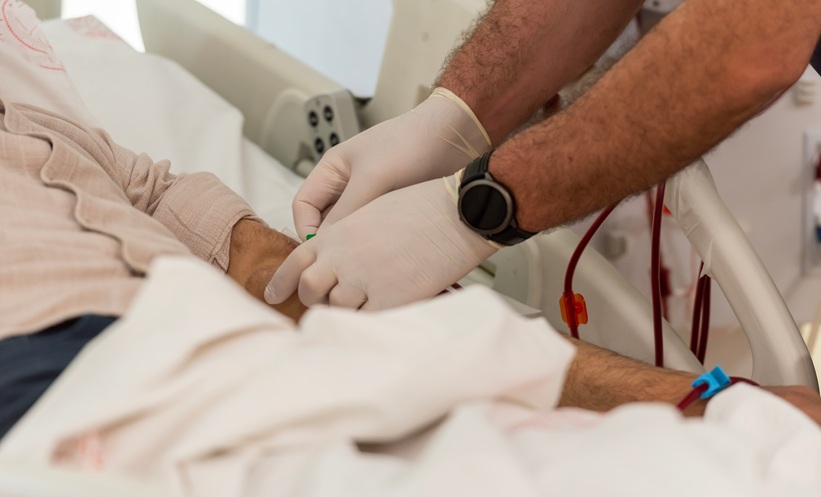
2. Figure 1: this shows positive wrist sign. The patient was asked to grip his wrist with his opposite hand. The thumb and fifth finger overlap, hence the sign is positive.4
Positive facial features of MFS that the patient had include: micrognathia, malar hypoplasia, and downward slanting palpebral features.
A transthoracic echocardiogram showed aortic root dilation with aortic diameter Z >2.
A literature review using Pubmed, Google Scholar, and Semantic Scholar did reveal associations with ADPKD and connective tissue disorders.
This appears to be one of the very rare cases of MFS with coincidental ADPKD.

Figure 1: Positive wrist sign.
DISCUSSION
ADPKD has been associated with increased risk of intracranial aneurysms5 and connective tissue disorders.6 ADPKD is the most common monogenic form of inherited kidney disease worldwide.7 It is a common cause of end-stage kidney disease. Growth of renal cyst size and kidney volume over time precedes GFR decline. Kidney volume assessment by imaging is used for prognostication purposes. There is currently no defined treatment to reverse cyst size. Strict blood pressure control was shown to reduce adverse outcomes,8 and a low-sodium diet plus maintaining a dilute urine (as tolerated) is recommended. More recently, antidiuretic hormone antagonists like tolvaptan have been shown to reduce growth or renal cysts and slow the rate of GFR decline.9 One study showed benefit in GFRs above 60 mL/min/m2.10 This patient’s most recent estimated GFR measurement was 120 mL/min/m2 per CKD-EPI equation.
MFS is a predominantly autosomal dominant disorder with rare case reports described as recessive inheritance.11 The clinical presentation is broad, with clinical features involving ocular, cardiovascular, musculoskeletal, and, in some cases, lung, skin, and nervous system abnormalities. Most patients with the typical Marfan phenotype harbour mutations involving the FBN1 gene. FBN1 mutations can also show milder phenotypes. The details of genotypes and phenotypes of MFS is beyond the scope of this review. The diagnostic criteria is based on the presence or absence of family history of MFS. Based on aortic root dilatation, ectopia lentis, and systemic score (Ghent classification), this case had MFS. Surveillance imaging is recommended every 6 months to look for aortic root enlargement, and is the plan going forward for this patient.
Overlap Between PKD1 and FBN1 Mutations
A co-occurrence between cystic kidney disease and Marfan-like features was first described by Booth et al.12
ADPKD is primarily due to PKD1 and/or PKD2 gene mutations on chromosome 16 (resulting in polycystin-1 and polycystin-2 abnormalities, respectively) while MFS is due to FBN1 gene mutation on chromosome 15 (which results in upregulation of TGF-β). Cerebral vascular aneurysms have been reported with ADPKD.5 Aortic aneurysms also have been reported with ADPKD at a rate of 7.3-times greater than chance association alone.13 A possible reason for higher association of aneurysms and vascular problems in ADPKD could be advanced kidney disease itself. However, there are hypotheses that connective tissue defects could contribute to the pathogenesis of ADPKD,14 and that ADPKD by itself could be a disorder of connective tissue.
A study in 1993 by Samlo et al.6 studied patients with ADPKD and overlap connective tissue disorders. It suggested that coexisting connective tissue and vascular issues in ADPKD could be an ‘extension’ of PKD1 phenotype. One possibility discussed was that the disorders are produced by independent, but genetically linked, mutations. Hateboer et al.15 carried out a study to look at the genetic linkage between these two diseases.
Their conclusion was that these diseases do not appear to be genetically associated, and co-occurrence previously has been reported as a chance event. Per the original paper, which was carried out in Europe, the prevalence of ADPKD then in Europe was 1 in 1,000, and the rate of MFS in the UK was 1 in 14,000. Therefore, per their calculation, the chance of co-occurrence is 1 in 14 million.
There are reports that PKD1 mutations can cause a TGF-β upregulation, which exacerbates the MFS phenotype. A study was conducted by Liu et al.16 in mice, to investigate a potential genetic interaction between PKD1 and FBN1 mutations. Mice heterozygous for PKD1 were matched with those heterozygous for FBN1. Double heterozygotes for PKD1 and FBN1 mutations showed severe aortic abnormalities compared with those without dual mutations. That study went a step further and stained the aortic vascular walls. There was increased aortic staining for connective tissue growth factor in double heterozygotes. TGF-β signalling was increased in vascular smooth muscle cells isolated from PKD1 and FBN1 heterozygotes.
Aortic vascular abnormalities occurring in the setting of PKD1 mutations in isolation without other abnormalities have not been reported.17 Thus, the presence of aortic vascular abnormalities in patients with ADPKD should prompt clinicians to further evaluate patients for co-occurrence of other connective tissue diseases.
Studies have shown that angiotensin 2 induced the TGF-β pathway in ADPKD and aneurysm formation.18,19 Angiotensin receptor blockers were potent inhibitors of this pathway in FBN1 mutant mice, which could play a significant role in their management.
CONCLUSION
MFS and ADPKD are disorders that result in systemic connective tissue symptoms that can appear to overlap. However, there does not appear to be an actual genetic association between these two diseases.
Specifically, aortic vascular issues in those with ADPKD should prompt clinicians to evaluate patients for other aetiologies. More research is needed to identify the common signalling pathways in these rare disorders. Some studies described above have suggested that a more ‘muted’ form of FBN1 mutations in the presence of PKD1 mutations can increase the characteristics of MFS. Strict blood pressure control (preferably with angiotensin receptor blockade) and close surveillance for vascular abnormalities will be an integral part of management. This is especially important as the severity of vascular abnormalities can be more pronounced when the diseases co-exist.
This patient’s blood pressure was below 120/70 without medication, and they are being followed closely for surveillance of aortic disease. This report has some limitations because of the lack of specific genetic testing due to the patient’s financial constraints, as they have no medical insurance. This also limited some follow-up testing. Images for aortic root dilatation were not available.
However, the author can confirm that the patient has ADPKD, based on sonological criteria and family history, and MFS confirmed by clinical testing. The teaching point for nephrologists is that ADPKD and MFS can overlap. However, based on current data, this is purely a chance event, but further research is needed to identify common genetic pathways that could cause overlap between both disorders. This review of existing literature does show that ADPKD and connective tissue disorders can present together.