There is some controversy regarding left ventricular ejection fraction (LVEF) improvement after chronic total occlusion (CTO) recanalisation. While a meta-analysis of observational studies reports an average improvement of 4.4%,1 there is only one randomised clinical trial to demonstrate a benefit in the subgroup of patients with CTO of the left anterior descending artery.2
However, should LVEF improvement be expected after a single-vessel percutaneous coronary intervention (PCI)? Does it matter if this PCI is performed over a CTO regarding LVEF improvement? Maybe not. Moreover, we know from several registries that ≥50% of CTO patients have normal LVEF, so no relevant LVEF improvements might be expected in those patients.
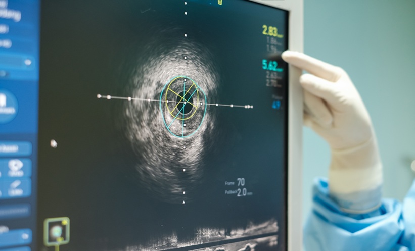
Longitudinal strain (LS), which is measured by two-dimensional speckle-tracking and represents myocardial deformation, is easily quantified in a semiautomatic fashion. It has independent prognostic value in a wide array of cardiac diseases, and might identify viable myocardium in ischaemic heart disease. We explored whether several LS parameters could be superior to conventional echocardiographic parameters in assessing LV function in CTO patients. Taking a 16-segment LV model from 13 consecutive, successful CTO PCI patients, a total of 208 segments were assessed.
No changes in global LVEF, telediastolic diameter, or mitral peak velocity of early filling to early diastolic mitral annular velocity ratio (E/E’) were detected at 1 or 3-month follow-up (56.0±6.0% at baseline versus 59.0±6.0% at 3-month follow-up; p=0.2; telediastolic diameter 54.8±26.0 mm versus 59.7±25.0 mm; p=0.6; and E/E’ 12.6±4.3 versus 11.8±3.8). Deformation parameters from CTO dependent or nondependent segments remained unchanged at 1-month follow-up (nonsignificant). At 3-month follow-up, the strain of CTO-dependent segments improved significantly: S-epicardial Δ -2.6±5.2; p<0.001; S-mesocardial Δ -2.3±5.2; p<0.05; and S-endocardial Δ -2.1±5.6; p<0.05. However, strain rate (SR) in CTO-dependent segments had no differences at 3-month follow-up: SR Δ -0.05±0.42; p=0.353. As expected, CTO nondependent segments did not change significantly.
In conclusion, this pilot study suggests that regional LS improves in CTO-dependent segments after successful PCI, while global LVEF, E/E’, and SR remain unchanged. Changes were not present after 1 month but became evident after 3 months. The discussion after the abstract presentation opened several questions to be answered with a greater number of patients and further follow-up. Future implications are the potential for LS to predict viability with incremental value over cardiac magnetic resonance, clinical implications of regional LV function improvement (such as better exercise tolerance), and clinical outcomes.