Author: Louise Rodgers, Editorial Assistant
On Day 2 of EuroPCR 2021, discussants from around the world took to their screens to join Jean Fajadet, PCR Vice-Chairman, and his panel, live from their studio in Paris, France. The aim was to understand how to adapt treatment strategy to the underlying anatomy in a case of left main bifurcation percutaneous coronary intervention.
THE CASE
The session began with a description of the case of a non-diabetic male patient with a history of a percutaneous coronary intervention to re-open a chronic total occlusion of the proximal mid-left anterior descending artery (LAD). In 2018, the patient presented with angina and a positive stress test at the Institute Cardiovasculaire Paris Sud, Massy, France. A coronary CT angiogram (CCTA) from 2018 displayed a lesion on the left main, a large circumflex artery, and obtuse marginal (OM) branch. The LAD was patent and there was an occlusion at the origin of the major septal branch. The team in Massy observed collateral flow from the dominant tricoronary artery with a distal LAD and second diagonal branch. The team performed the chronic total occlusion with two wires: one in the LAD and one in the diagonal branch. The operation was a success.
The patient recently presented again as symptomatic with recurrent angina and a positive stress test. On evaluation with a non-invasive CT an open, patent, dominant right coronary artery was visible, with no plaque present on the mid-segment and no critical stenosis. On the left, the OM branch appeared patent with slight black calcified plaque. The left main was severely diseased, with an eccentric lesion and an LAD with patent stent on the proximal mid-LAD. Immediately, it was noted that what they were viewing was a trifurcation of the left main and the lesion was distal, taking an origin from the LAD and circumflex artery. The most recent laboratory results came back normal and the patient is currently on dual antiplatelet therapy, β-blockers, calcium antagonists, and statin therapy. The most recent CCTA, presented to the EuroPCR audience, showed a short, discreet, distal left main lesion involving the large LAD, which was totally patent, and a large circumflex artery. The fractional flow reserve, performed in the distal LAD, came back positive (0.78) and the patient’s SYNTAX score was 21.
THE DISCUSSION
The task of the panellists was to discuss what their approach would be to such a case of a trifurcation. Fajadet began by addressing cardiac surgeon Thomas Modine, Centre Hospitalier Universitaire de Bordeaux, Bordeaux, France, to ask how he would handle this particular case at his centre. The case, in Modine’s opinion, perfectly illustrated the need for communication between cardiologists and cardiac surgeons. Recurrence of angina is a potential outcome regardless of how effective the first treatment or surgery was, given the evolutionary nature of the disease. Due to the success of the first surgery, the patient’s good quality arteries, and the patient’s age, he believed future surgery to be very possible. He concluded that the case was a good indication for both medical and surgical treatment and that at his centre, he would have most likely performed a memory graft to cover all the left branches.
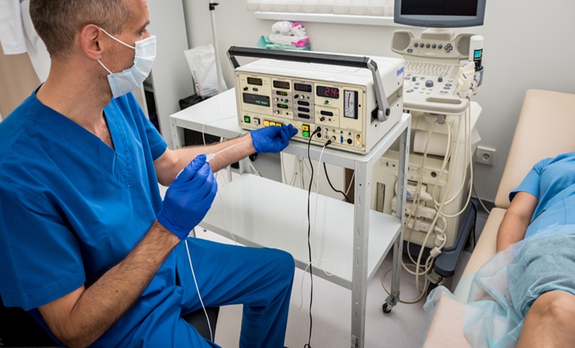
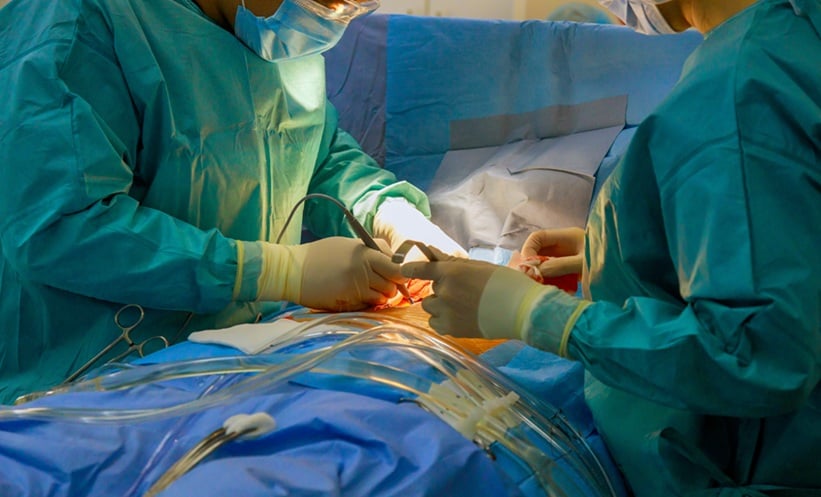
The screens turned to the live surgical case at the Institut Cardiovasculaire Paris Sud, Massy, France, where the surgeons were operating on the case in discussion. The team used a 7 French guiding catheter for extra support and backup. Three wires were inserted: one into the circulation, one into the intermediate branch, and one into the LAD. Imaging guidance had already been performed and the pullback with optical coherence tomography had been completed.
The team raised the question as to whether it technically was a trifurcation due to the gap between the ostium and the ostium circumference of the OM branch. The diameter of the LAD was approximately 3.5 mm and there was ostial stenosis of the LAD, which became visible to the audience on the CCTA. The surgeons attempted an optimal viewing angle, crucial for bi- and trifurcation cases.
After viewing the live case in Massy, the discussion turned to the hub in South Africa, where Farrel Hellig, Sunninghill Hospital, Johannesburg, and Mpiko Ntsekhe, Groote Schuur Hospital and University of Cape Town, Cape Town, and colleagues, were asked with regard to their particular strategy.
Based on the optical coherence tomography images from the live operation, the team in South Africa came to the consensus that they would go for a provisional single-stent strategy from the left main ostium into the proximal LAD, followed by a proximal optimisation technique, and thereafter assess those results to determine whether double or triple kissing inflation was required. They concluded that kissing inflation into the intermediate branch was not necessary; however, this would need to be assessed to determine whether additional stenting was required. The use of a single stent was regarded as the most likely outcome. In their final remarks for the case of bi- or trifurcation, most of the team members agreed that they would be comfortable with the use of a 7 French and that the use of three wires was optimal; even after having seen that it was not technically a true trifurcation, three wires was the safest option in order to protect all three branches.