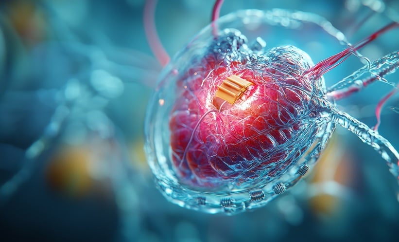
A THREE-DIMENSIONAL (3D) model of a human left heart ventricle has been successfully bioengineered by researchers at Harvard University, Cambridge, Massachusetts, USA. The new creation was built with the aim of providing a device that can be used to test drugs, study disease, and develop patient-specific treatments for arrythmia and various other heart conditions.
The heart model, which has been >10 years in the making, consists of a nanofibre scaffold that has human heart cells scattered within it. This acts as a 3D template that provides the cells with the guidance they need to form the ventricle chambers that beat in vitro. This model is a novel method for researchers to study the function of the heart using the same clinical tools as would be used on a real human heart, such as pressure–volume loops and ultrasound.
The nanofibre scaffold was developed using a platform known as pull spinning, which involves a high-speed rotating bristle that pulls a droplet of polymer solution from a reservoir into a jet. The polymer spirals under speed and, once solidified, detaches from the bristle and moves towards a collector. The ventricle consists of a combination of biodegradable polyester and gelatine, obtained using a bullet-shaped rotating collector. As the collector spins, it allows all of the fibres to align in the same direction.
The researchers used either rat myocytes or human cardiomyocytes from induced stem cells to culture the ventricle. After 3–5 days, a thin wall of tissue developed over the scaffold and the cells were beating in synchronisation. Using this newly developed model, the researchers studied the pressure and volume of the heartbeats through insertion of a catheter and could control and monitor calcium propagation. They were also able to expose the ventricle to isoproterenol and assess the changes in beat rate, as well as mimic, and subsequently assess, a myocardial infarction by pushing holes into the fabric of the model.
To ensure they could continue using the heart model, the researchers built a self-contained bioreactor with separate chambers, optional valve inserts, optional ventricular assist capabilities, and additional access points for catheters. In doing so, the team were able to keep the model stable and continue monitoring it for 6 months.
“The long-term objective of this project is to replace or supplement animal models with human models and especially patient-specific human models”, said Luke MacQueen, Harvard University, first author of the study. In the future, the researchers hope to use pre-differentiated stem cells directly from patients to create these ventricles, allowing for personalised treatment techniques and a higher throughput production of the tissue.


