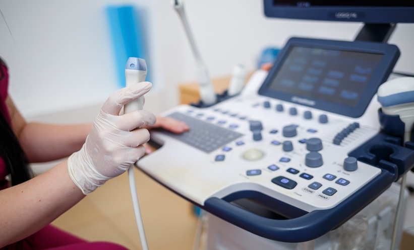
ULTRASOUND radiomics combined with machine learning showed potential for differentiating diabetic kidney disease (DKD) from non-diabetic kidney disease (NDKD) in patients with Type 2 diabetes mellitus (T2DM), according to a retrospective multicentre study. The findings suggested that integrating renal ultrasound radiomic features with eGFR could provide a non-invasive approach to preliminary disease differentiation.
The study included patients with T2DM who underwent renal biopsy across three centres. Biopsy findings were used to classify patients as having DKD or NDKD, with overlapping lesions assigned to DKD. Renal ultrasound images were analysed using regions of interest to extract radiomic features, while clinical predictors were selected using statistical and machine learning methods.
Machine Learning Model Shows Variable Performance
The researchers evaluated support vector machine, k-nearest neighbours, random forest and XGBoost models using radiomic features alone or alongside clinical variables. Serum creatinine, eGFR, albumin and low-density lipoprotein cholesterol were identified as significant variables in univariate analysis, with eGFR ultimately selected as the clinical predictor.
The integrated XGBoost model combining ultrasound radiomics with eGFR achieved an area under the curve: 0.991 in the training cohort; 0.895 in the internal validation cohort; 0.721 in the external validation cohort. Corresponding F1-scores were: 0.939; 0.815; 0.714.
The radiomics-only model performed well in the training cohort but showed lower performance in the validation cohorts. DeLong’s test indicated that XGBoost performed better than several comparator algorithms in the training and internal validation cohorts, while differences between models were not significant in the external validation cohort.
Potential Role in Preliminary Screening
The findings indicated that ultrasound radiomics could complement routinely available clinical information when distinguishing DKD from NDKD in patients with T2DM. The authors proposed that the integrated model could provide supportive information for preliminary screening in primary-level hospitals and nephrology departments.
By combining renal ultrasound imaging with eGFR, the approach could potentially assist individualised assessment and inform decisions about renal biopsy. However, performance in the external validation cohort was lower than in the training and internal validation cohorts, highlighting variation in model performance across cohorts.
Overall, the study supported further evaluation of ultrasound radiomics and machine learning as non-invasive auxiliary tools for differentiating kidney disease in patients with T2DM.
Reference
Chen W et al. Ultrasound radiomics-based machine learning models for differentiating diabetic kidney disease from non-diabetic kidney disease in type 2 diabetes. BMC Nephrol. 2026:DOI:10.1186/s12882-026-05353-7.