Written by James Coker | Reporter, European Medical Journal | @EMJJamesCoker
![]()
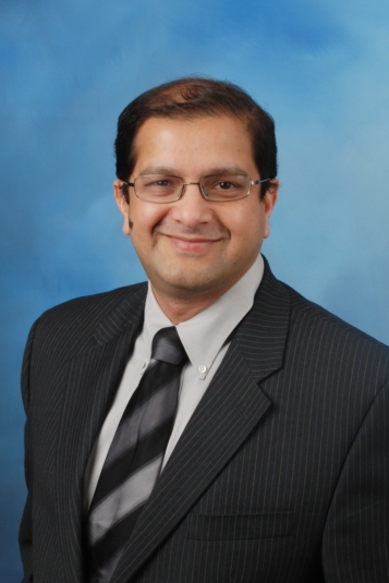
 During the prestigious Neuro Convention conference, which took place at the ExCeL, London, UK from 6th–7th June 2018, the European Medical Group were delighted to speak to several of the noted speakers in attendance. One of these was Dr Sanjay Prabhu, who gave an illuminating presentation entitled ‘Evolving Role of Machine Learning in Brain Tumour Imaging’ in which he discussed the growth of machine learning in the acquisition and interpretation of imaging studies performed in children with brain tumours. There is perhaps little surprise to learn that the paediatric neuroradiologist has a keen interest in the use of emerging technologies in this field. These include machine learning, artificial intelligence (AI), 3D printing, and augmented and virtual reality (AR and VR). We discussed these areas in great detail, in particular the ways in which such innovations have been incorporated into the day-to-day work of clinicians, including by Dr Prabhu in his clinical role as a Staff Pediatric Neuroradiologist at Boston Children’s Hospital. As will be displayed in this article, these exciting advancements are enabling rapid improvements in the diagnosis and development of precision therapies for neurological abnormalities, such as epilepsy, as well as for many other therapeutic areas.
Inspiration
Dr Prabhu was first inspired to work in radiology after experiencing the crucial role radiologists play in diagnosing conditions. This persuaded him to train in this field and ultimately specialise in paediatric neuroradiology, viewing the brain as an area in which there is wide scope for improvements in the diagnostic process.
“Now I practise paediatric neuroradiology alongside my work in AI and 3D printing, so it gives me two parts: a clinical hat where I work as a diagnostic radiologist and also work on 3D printing and AI all in one. It is a fascinating area because there’s so many things we don’t yet know; so there’s a lot of scope for improvement,” he explained.
Focus on Paediatrics
The focus on paediatric neurological disease is due to the huge potential for using technologies to develop very specific, personalised therapies for these patients. “I would say the brain is arguably one of the easiest areas to bring out like AI or machine learning because you have quite a stationary structure compared to other parts of the body,” elucidated Dr Prabhu. “Not only the brain is more stationary, but one also has two sides to compare and the fascinating thing about the paediatric brain is that it changes over time from a fetus to an 18-year-old. It undergoes rapid change over 18 years and it’s fascinating to see those changes and be able to develop applications that can diagnose conditions early on and predict what’s going to happen in later life. This has the potential to enable tailoring treatments for the child even before the disease shows itself.”
Advancements in Brain Imaging
Over the course of his career, Dr Prabhu has witnessed major advancements in brain imaging technology. He described how previously thick slice imaging was the norm, but technology has now advanced to the point where new types of imaging are enabling increasingly detailed insights into the brain. One example of this is isotropic and volumetric imaging and use of multislice scanners rather than those that are single slice. “Advances have moved so quickly; it’s a fascinating era to be in,” he added.
These advances in imaging have enabled a greater understanding of the mechanisms and genetic causes of many neurological conditions. Dr Prabhu explained how older scanning technologies would often fail to reveal any abnormalities that were present in conditions, such as autism and seizures, preventing timely diagnosis and appropriate treatment being initiated. Now, it has become much easier to pinpoint the cause of a condition and target treatment precisely for that patient. “In patients with drug-resistant seizures, for example, previously you had to take out large parts of the brain to get rid of the seizure focus, akin to cutting down the forest to get rid of a single leaf. Now you can direct your therapy to a particular spot where the lesion is,” commented Dr Prabhu. “We now have a robot in Boston Children’s Hospital that we can use to restrict our surgical procedure to get just that one small area and reduce the collateral damage around it. That is amazing, and all this has happened in the last 10 years or so.”
Rise of Machine Learning
The growth of machine learning in recent years is also facilitating personalised patient care. Machine learning allows all the new data that are becoming available via the use of updated technologies to be collated, enabling a detailed analysis of the problem. This means that rather than relying on the experience of a single surgeon, information gained from many previous cases can be brought together; this helps the surgeon pinpoint the treatment far more accurately. “Machine learning is growing because imagery is much better, genetic information from patients is much better, and you can collate all this only if you have the mechanism to bring all the data from disparate systems together. Once you get them all together and you look at them as a whole, you start seeing patterns, and that helps you direct therapies, helping the clinician and the patient,” stated Dr Prabhu.
The paediatric neuroradiologist also views machine learning as being important for freeing up the time of radiologists. For example, this technology has the ability to analyse lots of images of a single patient’s anatomy over multiple timepoints, and therefore outline changes that have occurred over time in a clear graphical format. The radiologist can therefore be alerted to any substantial changes that could necessitate treatment without having to pore over the images themselves, which can be difficult and time-consuming.
Debate About Mass Use
Despite the massive benefits of this technology, Dr Prabhu cautions that machine learning and indeed AI applications are not yet at a stage where they can be applied universally, arguing that simply uncovering new data and developing algorithms is not enough. He stated that these apps should be vetted and tested by experienced radiologists in the real world to ensure that they work properly in all kinds of scenarios. “There’s a debate ongoing right now about when to get the AI apps to the desktop and whether clinicians other than radiologists have access to it. Also, we are debating whether patients should be allowed access to it?” he said. “I do not believe we are at a stage where these machine learning applications are available for mass use; I don’t think patients should be able to run their scans with the AI apps out there until they have been vetted out thoroughly.”
Growth of 3D Print
Another area that Dr Prabhu is heavily involved in is 3D printing. There is a world-renowned SIMPEDs3D Print programme at Boston Children’s Hospital, where he is Clinical Director. He explained that 3D printing has now become part of the usual procedure for surgeons about to undertake various types of complex operations at this institution and others, including for spinal deformities, craniofacial abnormalities, and tumour resection. A 3D print is taken of the patient prior to the operation, enabling the surgeon to essentially practise the surgery before the real event, preparing them for any variations and complications that could occur in that particular patient, thus ensuring that the actual operation goes smoothly. The prints are also very helpful for rare conditions that the surgeon may not have previously experienced. “It’s like taking a swing in a game of golf before taking the tee shot: the surgeon gets the chance to practice before going into the operating room (OR),” he explained. “So they learn all the variations in their patient before going in to the OR and it reduces complications, sedation time, and bleeding time in the OR.”
Dr Prabhu described how this use of 3D printing is gaining greater traction, despite initial scepticism from some quarters; he gave an example of an experienced spine surgeon colleague who now uses it for every operation he undertakes despite being very unconvinced of its usefulness to begin with. Clinical trials displaying the efficacy of the technique in terms of reducing complications in surgery is now growing, and Dr Prabhu has himself been involved in many of these.1,2 He believes that this increasing evidence will only lead to greater uptake by surgeons, especially in the context of reduced printer and material costs. However, he acknowledged that in certain therapeutic areas, such as oncology, the quality and speed of the prints needs to be improved before 3D printing can be relied upon as a valuable tool.
Future Potential
Nevertheless, he sees exciting possibilities for 3D printing in the future, particularly if it can be utilised in combination with other innovative technologies, which should assist surgeons with their procedures even more. He elucidated: “The next wave will be tying 3D printing with outward reality and virtual reality. So then you have this second layer of mixed reality: 3D print plus virtual technology, and synergy between these.”
In terms of future trends, Dr Prabhu firmly believes that there will soon be some very exciting developments in the fields of mixed reality, VR, and AR, and as alluded to above, these advances will be complementary to 3D printing in the OR. This should raise the visual capabilities of surgeons to new heights. While there is currently a certain amount of reluctance to use these technologies because of impracticalities of wearing headsets during operations, new innovations such as the updated Google Glass could help overcome such obstacles.3
EMJ Neurology 6.1
It was great to speak to Dr Prabhu about his passion for incorporating innovation and technology in healthcare, particularly in his own field of neuroradiology. The use of technology to diagnose and treat neurological conditions was a key feature of the presentations made during the 4th Congress of the European Academy of Neurology (EAN), which was held in Lisbon, Portugal from 16th–19th June 2018. This prestigious event will be reviewed in the upcoming edition of the EMJ Neurology eJournal, published shortly on 7th August. The journal will also include abstract reviews, interviews, and peer-reviewed papers; you can subscribe here for free to receive instant news of this publication as well as all our future content in the field of neurological health.
REFERENCES
- Karlin L et al. The surgical treatment of spinal deformity in children with myelomeningocele: the role of personalized three-dimensional printed models. J Pediatr Orthop B. 2017;26(4):375-82.
- Rogers-Vizena CR et al. Cost-Benefit Analysis of Three-Dimensional Craniofacial Models for Midfacial Distraction: A Pilot Study. Cleft Palate Craniofac J. 2017;54(5):612-7.
- Lamkin P. Google Glass Could Make Comeback In AR Revolution. 2018. Available at: https://www.forbes.com/sites/paullamkin/2018/02/26/google-glass-could-make-comeback-in-ar-revolution/#34f95afe23a6. Last accessed: 19 July 2018.


