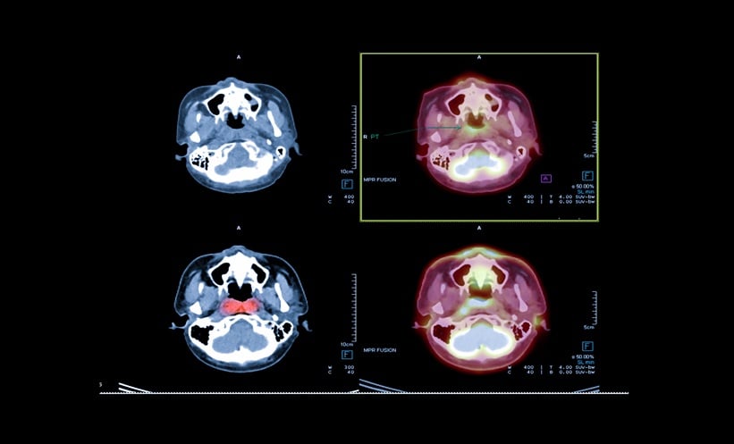
TARGETED PET imaging for glioma reliably distinguishes recurrent malignancy from radiation necrosis with strong clinical precision. In neuro-oncology practice, conventional magnetic resonance imaging frequently encounters diagnostic ambiguity when post-treatment tissue alterations, such as pseudoprogression, closely mimic tumor recurrence. Findings from the prospective registrational trial NCT04044937, supported by real-world validation cohorts, demonstrate that the amino acid tracer floretyrosine F 18 provides the metabolic differentiation necessary to resolve this critical clinical challenge in adult and pediatric patients.
Clinical Validation of PET Imaging for Glioma
The prospective multicenter study evaluated 127 patients presenting with suspected recurrent or progressive intracranial neoplasms following surgery, radiation therapy, or chemotherapy. Three blinded, independent nuclear medicine readers assessed the scans against a multidisciplinary composite reference standard combining serial imaging, clinical outcomes, and histopathology. The positive percent agreement of PET imaging for glioma in detecting true recurrence ranged between 70% and 83% across readers, while the negative percent agreement reached up to 64%. Among the 11 pediatric patients analyzed in the trial, positive percent agreement reached 83%. Complementary data from a retrospective cohort of 254 patients demonstrated positive percent agreement of 66% to 72% and negative percent agreement of 53% to 61%, confirming reproducible diagnostic performance across varied clinical environments.
Biological Mechanisms and Practice Implications
Unlike glucose-based imaging modalities that suffer from intense physiologic background uptake in normal brain tissue, floretyrosine F 18 exploits Large Neutral Amino Acid Transporter 1 and 2 mechanisms. Because these amino acid transporters undergo marked upregulation in glioma cells, the tracer achieves substantial tumor-to-background contrast that enables precise visual and semi-quantitative evaluations. Clinicians utilize target-to-background ratios to differentiate active malignancy from treatment-related tissue injury, with thresholds of mean ratio at or above 2.0 and maximum ratio at or above 2.3 indicating disease progression. The diagnostic procedure demonstrated a favorable safety profile across 382 evaluated patients, with headache representing the only adverse reaction occurring in at least 0.5% of individuals. Integrating PET imaging for glioma into post-treatment surveillance equips neuro-oncology teams with actionable diagnostic clarity, mitigating unnecessary invasive biopsies while directing timely salvage interventions.
Reference
Hope TA et al. Fluoroethyltyrosine for evaluation of intracranial neoplasms. ClinicalTrials.gov. 2026;NCT04044937.
Featured Image: Samunella on Adobe Stock.