BACKGROUND AND PURPOSE
Thyroid disease affects 42 million people in India and among these 12% have a thyroid nodule. Furthermore, India and the USA have both recorded a steep rise in thyroid cancer incidence during the last decade.1 Though the sensitivity of ultrasound for detection of a thyroid nodule is high, it shows a dismal performance for differentiation between benign and malignant nodules.2 Moreover, the not so satisfactory performance of thyroid imaging reporting and data system (TIRADS) in characterisation of thyroid nodules has been substantiated in a meta-analysis by Wei et al.; the accuracy of TIRADS stratification showed a pooled sensitivity and specificity of only 75% and 69%, respectively.3 The American College of Radiology (ACR) TIRADS was developed in 2017 for better characterisation of thyroid nodules.4 While ACR TIRADS 1 and 2 are certainly benign nodules and TIRADS 5 are definitely malignant, TIRADS 3 and 4 nodules are unfortunately in a rather grey zone. In this context, various investigators have explored the application of shear wave elastography (SWE) for better characterisation of thyroid nodules.5-8
Therefore, the aim of the present study was to assess the diagnostic performance of established SWE values in evaluating and predicting malignancy in ACR TIRADS 3 and 4 suspicious thyroid nodules, using cytology and histopathology as the gold standard. The secondary purpose was for the authors to determine their own SWE value cut-offs in definitively characterising thyroid nodules as benign or malignant.
MATERIAL AND METHODS
This was an institutional review board-approved prospective study, standardised by using the same ultrasound equipment (Siemens Acuson S3000TM; Siemens Healthineers, Erlangen, Germany) and the same two radiologists performing the ultrasound each time (first and second authors). The patients first underwent conventional ultrasound and the nodules were classified according to ACR TIRADS system. Sixty consecutive patients with TIRADS 3 or 4 lesions were recruited for the SWE study. In patients with multiple nodules, the single most suspicious nodule was interrogated. Lesions were classified into benign or malignant using the previously established SWE cut-off value of 85.2 kPa, with higher values being considered as malignant.5 Pathology diagnosis obtained either by fine-needle aspiration cytology or biopsy was considered as the gold standard. Statistical analysis was performed for determining sensitivity, specificity, negative predictive value (NPV), positive predictive value (PPV), and diagnostic accuracy of SWE, based on established cut-off values. SWE values obtained from the current cohort were correlated with final pathology diagnosis and retrospectively evaluated for construction of a receiver operating characteristic curve. A new cut-off value for SWE was then computed and proposed.
RESULTS
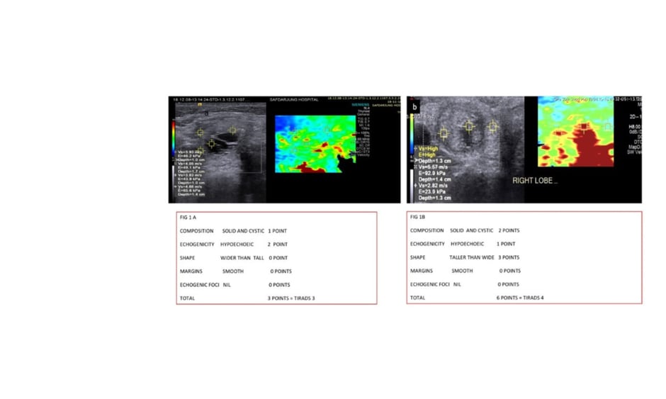
In evaluated nodules, ACR TIRADS classified 29 nodules as TIRADS 3 and 31 as TIRADS 4. Using established SWE cut-offs, 27 nodules were classified as benign and 33 as malignant (Figure 1). Fine-needle aspiration cytology and histopathology revealed 28 nodules as benign and 32 as malignant. Using established SWE cut-offs, sensitivity, specificity, PPV, NPV, and diagnostic accuracy were 81.3%, 96.4%, 96.3%, 81.8%, and 88.3%, respectively. The retrospectively determined cut-off value for SWE was 74 kPa, with sensitivity, specificity, PPV, NPV, and diagnostic accuracy of 96.9%, 85.7%, 88.6%, 96.0%, and 91.7%, respectively.
CONCLUSIONS
SWE using established cut-off values shows good diagnostic performance for a more definitive characterisation of TIRADS 3 and 4 nodules. The lower SWE cut-off obtained in our study differs significantly from that obtained in other studies (p<0.05) and shows a superior sensitivity, NPV, and diagnostic accuracy to the previous values. The authors propose that a SWE cut-off value of 74 kPa should be used in future studies, in view of patients’ safety towards avoiding repetitive biopsies. ■
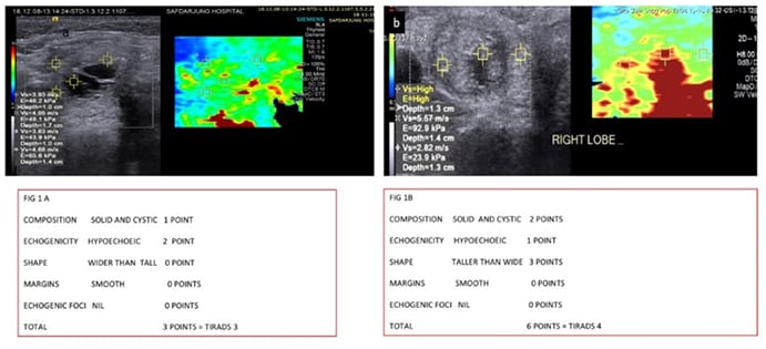
Figure 1: SWE in TIRADS 3 and 4 nodules. A) The SWE study in a 25-year-old female patient with a TIRADS 3 thyroid nodule. The SWE value was 65.6 Kpa, the biopsy and histopathology revealed a colloid nodule. B) The SWE study in a 27-year-old female patient with a TIRADS 4 thyroid nodule. The SWE value was 92.9 Kpa, the biopsy and histopathology revealed a papillary carcinoma. SWE: shear wave elastography; TIRADS: thyroid imaging reporting and data system.