
BACKGROUND AND AIM
CT has become an established practise in forensic imaging, in which this method is called
post-mortem CT. In contrast to clinics, to date only a small number of published scientific publications on protocol optimisation exist. Because the accuracy of radiological image data evaluation is dependant on the image quality, these studies are essential. Furthermore, compared to the examinations of living patients, motion artifacts and radiation dose do not have to be considered in the post-mortem field.1-8
The aim of this study was to evaluate the current institute standard of the Institute of Legal Medicine for a native cardiac post-mortem CT at the University Medical Center Hamburg-Eppendorf, Hamburg, Germany, in regard to image quality and whether or not optimisation of this protocol was achievable.
METHODS
For this purpose, a prospective study with five bodies was carried out in January 2019. The cadavers were scanned with five different heart protocols, which is the institute standard, and four protocols were developed for this study. The protocols deviated from each other in the scan parameters kVp, mAs, slice thickness, and increment (Table 1). Scan range was from the middle of the larynx to the phrenicocostal sinus. Each cadaver was stored in supine position, the arms were tied together using medical tape and were positioned over the head.
The protocols were assessed by a 5-point ordinal-LIKERT-scale questionnaire from four independent radiologists, without knowledge of the institute standard. Using Microsoft Excel and SPSS Statistics, the questionnaires were evaluated and then inter-rater reliability (Kendall´s coefficient of concordance) was calculated in order to evaluate the agreement between the assessors.
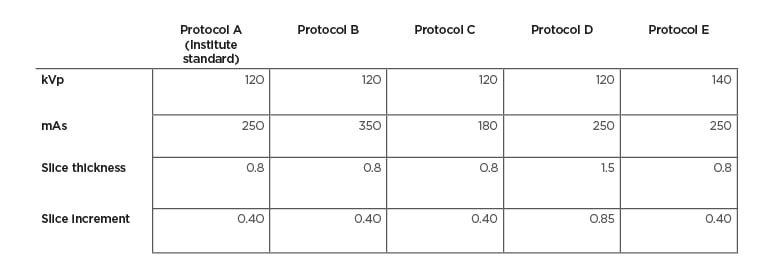
Table 1: Overview of all five cardiac scan protocols.
Protocol A is the institute standard, the other four protocols were created for this study.
RESULTS
The results show that the higher slice thickness protocol was rated higher in terms of image quality than any other protocol. Likewise, the two higher-dose protocols also performed better than the institute standard. The lower-dose protocol was rated as the worst. After calculation of the inter-rater reliability, an acceptable agreement between the raters could be assumed.
DISCUSSION
Despite the clear results, there are some limitations regarding this study; only a small number of cadavers were included and only adult cadavers were studied. In order to ensure a practical environment, the protocols must also be examined in children, infants, and adolescents. Furthermore, the protocols in the image data received by the raters were labelled and arranged in the same way in all cases, which is why a bias of the assessors cannot be ruled out. Additionally, the results did not allow for any evaluation of the protocols’ diagnostic value. For this, further studies are necessary. Future research should also clarify the question of whether a combination of a higher slice thickness with a higher dose can lead to a more enhanced image quality.


