BACKGROUND AND AIMS
PET/MRI scans can provide a significant advantage in the paediatric age group, because they are more radiosensitive than adults and more prone to radiation induced long-term adverse effects.1 Radiation exposure can be significantly reduced by up to 70% by replacing CT scans with MRI using hybrid PET/MRI scanners.2 Additionally, the higher sensitivity of solid state PET detectors in current PET/MRI scanners, and the possibility to extend PET acquisition times due to the simultaneous acquisition of PET and MRI, make it possible to further reduce the radiation exposure by decreasing the injected radiotracer activities.3,4 The aim of this study was to evaluate the effect of reduced injected tracer activities on the quantitative image metrics and the visual image quality in whole body 18F-FDG PET/MRI with time-of-flight capability in paediatric oncology.
MATERIALS AND METHODS
Seventy-seven oncological whole body PET/MRI examinations of 54 paediatric patients were analysed (standard injected activity: one-half dose [1.9 MBq/kg] and standard PET scan duration: 5 min per bed position). Lower activity PET images (one-third dose [1.2 MBq/kg] and one-quarter dose [0.9 MBq/kg]) were retrospectively simulated by truncating the originally acquired list mode data sets. In order to examine the influence of dose reduction on quantification, volumes of interest (VOI) were placed within normal organs: liver, mediastinal blood pool, bone marrow, psoas muscle, and bladder, and around FDG avid lesions. VOI positions and size were selected on the original data sets and copied to other simulated data sets for obtaining the same VOI size and localisation. Objective quantitative parameters were assessed by measuring the standardised uptake value (SUV) metrics: SUVmax, SUVmean, SUVvar, and SUVpeak, signal-to-noise ratio (SNR), and contrast to noise ratios (CNR) in each PET data set. Differences in quantitative parameters of simulated data were recorded as relative percentage changes compared to the original data. PET images were also evaluated visually for general image quality by using a 4–point scoring system (1: excellent, 2: good, 3: average, 4: inadequate/poor).5
RESULTS
SNR were significantly different among PET data sets (p<0.001) and showed gradually increasing image noise with decreasing doses. CNRmax and CNRmin did not show any significant differences among PET data sets (p=0.152 and p=0.259 respectively). The mean relative percentage changes in SUV metrics in physiological organs and in FDG avid lesions were found to be lower at one-third dose data set, compared to one-quarter dose data set. Lesion SUVmax and SUVmean values were significantly higher in one-quater dose data set, compared to the original and one-third dose data sets (p<0.05 for all). However, these SUV metrics did not show statistically significant difference between the original and one-third dose data sets.
While the mean visual score at one-quater dose data set was significantly higher compared to the original and one-third dose data sets (p<0.001 and p=0.001 respectively), there was no significant difference between the original and one-third dose data sets (Figure 1).
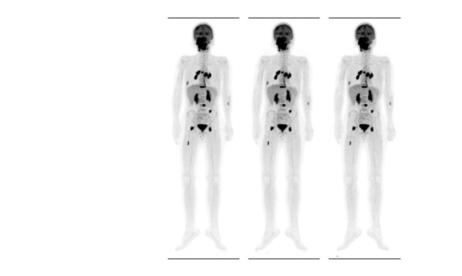
Figure 1: 11 year-old male with newly diagnosed embryonal rhabdomyosarcoma. Maximum intensity projection images of the patient for the original data set (1.9 MBq/kg), and simulated activities of 1.2 and 0.9 MBq/kg (from left to right). SNR were 10.2, 9.5, and 8.1, respectively. CNRmax values were 16.3, 15.3, and 15.2, and CNRmin values were 11.3, 10.3, and 9.8 respectively. In accordance with quantitative results, while there was no significant difference in the detectability of 18F-FDG avid lesions, image noise, and granularity increased with decreasing tracer activities.
CNRmax: Maximum contrast-to-noise; CNRmin: minimum contrast-to-noise; SNR: signal-to-noise.
CONCLUSION
The study’s quantitative and visual analyses showed that the reduction of injected activity to 1.2 MBq/kg can be feasible in paediatric oncological PET/MRI, with a smaller relative percentage change in quantitative parameters and with similar image quality to the original data set. The level of the lowest tracer dosing that is proposed in our study is lower compared to the lowest tracer dose limits recommended in the previous studies.6-8 ■


