Prof Nandita deSouza | Chairperson of the European Imaging Biomarkers Alliance – EIBALL
Institute of Cancer Research (ICR), London, UK
The Royal Marsden Hospital, London, UK
![]()
As the Chairperson of the European Imaging Biomarkers Alliance (EIBALL) for ESR, please could you tell us about the subcommittee and your role?
I took on a 3-year term to chair this committee, and we set out a mission statement and a 2-year roadmap in the first instance. The mission is: “To facilitate imaging biomarker development, standardisation, and promote their use in clinical trials and in clinical practice by collaboration with specialist societies, international standards agencies, and trials organisations to develop a network of excellence.” The 2-year roadmap has three pillars: 1) promoting biomarker usage in clinical trials; 2) setting standards for biomarker usage; and 3) educational activities that encourage the use of biomarkers in a standardised way within clinical trials. We are in the process of updating the roadmap for my third year. We maintain a policy of keeping the committee refreshed with new members to bring new insights and endeavours to achieve our mission.
Last year, EIBALL published a paper on validating imaging biomarkers for their use as decision-making tools. What was the reason behind the production of this paper?
The reason for making quantitative measurements is to be able to use the quantitation to drive clinical decisions regarding patient management. With any quantitation, it is important that the measurement is robust and reproducible wherever it is done, and that this does not vary because of variations in imaging hardware or software. We have to quality assure the measurement process and be aware of the confidence limits of the measurement. The purpose of the manuscript was therefore to discuss the necessary standards and set out recommendations for the imaging community when making quantitative measurements.
Collaboration is an essential part of scientific progression. Is the EIBALL subcommittee currently working on any projects with other committees or societies?
We work within an imaging community and collaboration is at the heart of what we do. Our biomarker inventory work is being done in collaboration with organ-specific specialist societies, for instance the European societies of urogenital radiology (ESUR), gastrointestinal and abdominal radiology (ESGAR), and breast imaging (EUSOBI). We work also with modality-specific societies and have members representing the European Society of Magnetic Resonance in Medicine and Biology (ESMRMB) and European Society of Hybrid Imaging (ESHI) on our subcommittee. We work with the European Society for Research and Treatment in Cancer (EORTC) to promote biomarkers within the clinical trials agenda. Finally, we have strong links with our North American partners from the Radiological Society of North America (RSNA) and have cross-representation on the EIBALL and their Quantitative Imaging Biomarker Alliance (QIBA) subcommittees. In particular, we are working with the QIBA metrology group to set standards for radiomic analyses and usage.
As co-director of the Cancer Research UK (CRUK) Clinical Magnetic Resonance Research Group at the Institute of Cancer Research (ICR), what are some of the projects that your research team work on?
During my 16 years at ICR, the MRI group have conducted a research programme with many separate but integrated aspects. We had funding from CRUK (2004–2019) to achieve a programme of work that spanned preclinical and clinical research. It covered a variety of topics, with a primary focus on tumour heterogeneity. We aimed to understand how tissue heterogeneity related to differences in biological behaviour, to differences in response to combination therapies (with a focus on targeted therapeutics), and eventually to outcome.
Your research is primarily focussed on the use of MRI to identify biological indicators to predict a patient’s prognosis and response to treatment. How has this field developed recently?
There has been a huge increase in the use of techniques such as diffusion-weighted MRI in tumour assessment, not just qualitatively, but also quantitatively, using metrics such as histogram parameters to characterise tumours and their response. Moreover, these data are available not just from dedicated regions, but with the advent of whole-body techniques, it has become possible to interrogate the entire skeleton, for example in patients with myeloma or skeletal metastases. Finally, the use of radiomics, which extracts data that is beyond visual perception from the images, will be a key methodology. It also lends itself to automated analysis, which is important as we move towards an era of artificial intelligence.
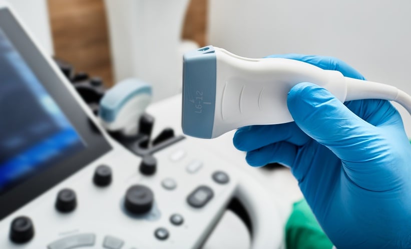
You have been noted as a pioneer for the use of endocavitary probes in MRI. How have these devices changed the approach to patient diagnosis and prognosis determination?
Endocavitary devices allow high-resolution imaging. They bring a receiver close to a region of interest and increase the signal in the region-of interest four to ten-fold. This allows a phenomenal improvement in image resolution, which is important when assessing early or small volume disease. We have used endorectal coils to assess prostate cancer, for which the technique has been particularly useful in following patients managed by active surveillance. These men have tumours often not identifiable on standard imaging, and just being able to confirm their slow progression has been advantageous. Another major area has been in cervical cancer, for which high-resolution images of the cervix are particularly critical for mapping the extent of small tumours before the patients are considered for fertility-sparing procedures such as trachelectomy.
You are credited for bringing high-intensity focused ultrasound (HIFU) treatment to the patients at The Royal Marsden. What are the benefits of this treatment?
HIFU is a very precise treatment that works by thermally ablating tissue at its focus, causing narrow linear burns. It has been used mainly for treatment of symptomatic fibroids. In cancer, it is used for treating pain from bone metastases by ablating nerve endings in the overlying periosteum. Together with University Medical Center Utrecht, we ran an international trial sponsored by Philips (Best, the Netherlands) and showed that pain and quality of life did indeed improve in these patients. This had been preceded by a randomised trial in the USA which showed the same outcomes. University Medical Center Utrecht are now running an international trial comparing HIFU to radiotherapy for treating painful bone metastases. We are part of this consortium, which is funded by a European Union (EU) H2020 grant. We have also done some pilot work using HIFU to treat symptomatic recurrences of gynaecological malignancy. The trial has recently closed, and we are in the process of analysing the data.
You have previously worked in the Women in Academic Medicine Committee (WAM) and are currently a member of the Athena SWAN steering group. Why is this an important issue for yourself and how can other healthcare professionals support women in academic medicine?
Supporting women to be successful is very important to me. Women are generally more reticent in how they present themselves, and they often lack the confidence to promote themselves in the same way as their male counterparts. Society judges success by male behaviours, and although these conceptions are slowly changing, it takes a long time and has to be constantly worked at. Interestingly, the three countries doing well in the COVID-19 crisis are all led by women who recognised the problem early and put the right measures in place quickly: Germany, New Zealand, and Taiwan. Actions here were more critical than political manoeuvring, and women can often be more practical. Women in academic medicine can need a helping hand at the top end of the ladder; more appointments to chairman positions and lead roles would inspire other women as they become role models and mentors.