This year, the European Society of Radiology (ESR) welcomed 17,262 participants from 122 different countries to their congress in Vienna, Austria, and online. After the online congresses in 2020 and 2021 due to COVID-19, and the congresses both in-person and online last year, ESR president Adrian Brady introduced this year’s congress as a tribute to the past presidents who dedicated themselves to achieving success in difficult times.
After a few disruptive years, during which the ESR had to show their innovation and willingness to change in order to ensure that their mandates and responsibilities were still fulfilled, the ESR has tried their best to create a safe environment in which the standard of professional and educational content remains high. Brady began the opening ceremony by thanking everyone who dedicated their time to making this congress a success, including all those involved in planning the congress, but also all ESR members.
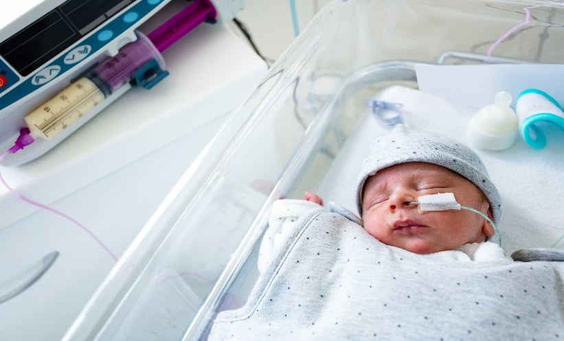
Brady then introduced the theme for this congress, ‘The Cycle of Life’. This theme aimed to bring attention to the importance of the role of radiology and radiography at all stages of life, from before it begins until after it ends. Numerous sessions throughout the programme were given this tag, so that participants could easily spot those sessions that cover a specific stage in life, or unusual applications of techniques. This theme allowed for a programme filled with unusual topics such as forensic imaging; the use of imaging in palaeontology and archaeology; the investigation of artworks and musical instruments; and the use of radiology in the generally healthy, for example through sport applications.
As is tradition, ESR chose one ‘in focus’ group to be given particular attention at the congress, trainees. While the congress features materials suited for practitioners at any stage of their career, in addition, the ESR added emphasis to areas that would interest trainees, including work-life balance, career development, mentoring, diversity, and equality and inclusivity. At the opening ceremony Brady gave advice to trainees in the audience, starting with the importance of accepting imperfection and trying to learn and strive to be the best you can be, while being kind to yourself and your colleagues when you or they fail after having tried diligently. Brady highlighted that radiology is a rapidly changing specialty, explaining that much of what is taught today will be redundant by the time practitioners reach the middle or end of their career. Brady stressed the importance of innovation in this change, saying: “Technical innovation, if embraced in the right way at the right time, can lead to huge leaps in human endeavour.” Finally, Brady reminded trainees to be aware of changes. The COVID-19 pandemic has created distance between radiologists and their patients and referrers. Brady encouraged trainees and their seniors to show others the importance of their role, and to make themselves indispensable to secure the future of radiology.
At the opening ceremony, six awards were granted in recognition of outstanding, internationally-recognised achievements and leadership. First, ESR honorary membership was granted to Beatriz González Ulloa, Dei Diagnóstico Especializado Por Imágen, Zapopan, Mexico, for their work in the field of breast imaging; Bruce G. Haffty, Rutgers Cancer Institute of New Jersey, New Brunswick, USA, for their work in radiation oncology; and finally, James Griffith, The Chinese University of Hong Kong (CUHK), for their work in the field of musculoskeletal imaging. Three gold medals were also awarded to Sue Barter, Addenbrooke’s Hospital, Cambridge, UK, for their work in breast and gynaecological imaging and in clinical audit and governance; Christoph D. Becker, University of Geneva, Switzerland, for their work in abdominal, diagnostic, and interventional radiology; and Lorenzo Derchi, University of Genoa, Italy.
The EMJ team was delighted to be a part of this congress, and is looking forward to ECR 2024, which will take place in Vienna from 28th February–3rd March. Read on for scientific highlights, covering topics such as saving energy in radiology departments and MR-planimetry in multiple sclerosis, as well as interviews with ESR president Adrian Brady, and European Federation of Radiographer Societies (EFRS) president Andrew England.
AI: Tacking Dataset Deficiencies in Brain Metastases
RESEARCH investigating the merit of medical artificial intelligence (AI) has previously suggested that AI is of little use in clinical practice due to poor quality datasets. The Stanford BrainMetShare Dataset, the only available dataset for brain metastases, is limited to 156 cases, focusing on just six primary tumour types. Work published attempting to predict tumours from MRI again only focuses on six of the most common primary tumour types. Thus, these datasets are inadequate as “many of the rare cases are regularly neglected,” noted Quirin Strotzer, Department of Radiology, University Hospital Regensburg, and Department of Neuroradiology, Medbo District Hospital Regensburg, Germany, who called for higher quality, diverse datasets in medical AI during their presentation at the European Congress of Radiology (ECR) 2023.
Strotzer and team recruited patients between 2003 and 2021 with cerebral metastases that had tumour histology and pre-therapeutic MRI data (T1, T1CE, T2, FLAIR) available. 3D T1 post-contrast MPRAGE data was also utilised where possible (87.6% of patients). Clinical parameters were recorded for each patient, including gender, age, Eastern Cooperative Oncology Group (ECOG) performance status, BMI, histology, survival, extracerebral metastases, and therapy. MRI data was then collected, and a bias correction was applied before co-registering the different images and applying an intensity normalisation. Based on a U-Net trained on brain tumour segmentation (BraTS) data, three different segment labels were then applied and manually verified: oedema, non-enhancing tumour, and enhancing tumour.
The final dataset contained 231 patients with 647 metastases. Fifteen different MRI scanners generated images, with the vast majority originating from 1.5T scans (89%). Unlike previous datasets, 27 primary tumour types were included, highlighting that the metastases most frequently originated from lung adenocarcinoma (181 cases), followed by melanoma (119) and breast cancer (85).
To conclude, Strotzer and team generated a standardised, multisite, multi-vendor dataset of pre-operative MRIs of brain metastases. They described it as the largest, most heterogenous dataset available, with higher accuracy to medical reality. In future, they believe the dataset could be easily expanded to incorporate DNA mutation data.
Mammography Screening Using Artificial Intelligence
RETROSPECTIVE results from a mammography screening study have been presented at the European Society of Radiology (ESR), held in Vienna, Austria, in March 2023. The aim of the Section for Breast Cancer Screening at the Cancer Registry of Norway was to compare rates of cancer detection using two CE-marked artificial intelligence (AI) systems, using the same retrospective data set.
Breast cancer is the most common cancer for females in Norway, and 3,991 females were diagnosed with the disease in 2021. In the country, females between the ages of 50 and 69 are invited for a screening mammogram at 2-year intervals, with the aim of finding early-stage breast cancer, and preventing unnecessary deaths from the disease. All breast screens in Norway are independently interpreted by two radiologists who specialise in breast imaging.
In total, the study analysed 106,380 examinations, which were carried out between 2009–2019 in the county of Østfold in southeastern Norway. The data set was provided by BreastScreen Norway, part of the Cancer Registry of Norway, and the study used images taken by Siemens MAMMOMAT Inspiration (Munich, Germany).
Measures were interpreted by two AI systems, A and B; during this process, 645 screen-detected cancers and 173 interval cancers were found. System A selected 91% (587/645) of screen-detected cancers, and 43% (74/173) of interval cancers, and System B selected 84% (540/645) of screen-detected cancers, and 37% (64/173) of interval cancers, respectively. A and B agreed in 53% of examinations selected in this study. Researchers found that 93% of screen-detected cancers and 50% of interval cancers were selected by systems A or B.
Solveig Hofvind, Section for Breast Cancer Screening, Cancer Registry of Norway, Oslo, Norway, pointed out that the limitations of the study are that it was carried out at a single centre; it is unknown whether the location detected by AI represents the real location of the cancer; and the differentiation between missed versus true interval cancers.
In future, Hofvind added, the analysis of the study will be extended to include more centres across all of Norway’s regions. Researchers also want to look into the possible benefits of using two or more AI systems working concurrently, and the use of AI as a support in screen-reading, or to triage screen examinations. Any future prospective studies will be based on this analysis, which was carried out using retrospective data.
No Time to Be Idle in Radiology Departments
RADIOLOGY departments consume large amounts of energy, with a single CT scanner having the energy consumption of five four-person households. Over two-thirds of this energy is used when in an idle state, according to Manfred Tobias Meyer, Department of Radiology and Nuclear Medicine, University Hospital Basel, Switzerland, who presented these findings at the European Congress of Radiology (ECR) 2023.
Meyer and colleagues monitored approximately 60 medical imaging systems, including five CT, six MRI, two PET-CT, 12 radiography units, four angiography units, and seven ultrasound machines, as well as 80 picture archiving and communication system workstations, 165 personal computers (PC), six smart monitors/PCs, and 53 printers in their department. They were able to determine the devices’ online status by internet control message protocol echoes, requested in 15-minute intervals, which were presented on live dashboards.
The devices were tracked for 2 weeks in October and November 2022. Then, the project was announced in mid-November to raise awareness. They continued to track device usage to analyse changes in behaviour, and identified constantly running modalities that could be turned off.
There was a reduction in the number of devices left on during off hours. For example, the two PET-CT scanners are now being turned off over the weekend and overnight. In the next 4 months, one scanner saved 2,366 kW/hour of energy, equivalent to 520.53 CHF and 302.85 kg of carbon dioxide emissions. The projected savings are 72,337.3 kW/year, equivalent to 19,530.90 USD, and 9,259.0 kg of carbon dioxide.
However, there were limitations in this study, as devices with no network connection (e.g., most monitors) could not be tracked, and printers could not be reliably tracked because the network status remains on while in standby.
Despite this, idle devices present a significant opportunity to save on costs and energy, and raising awareness had a quantifiable effect on the turn-off rates of PCs, with estimated yearly savings in Meyer’s department of 13.9 four-person households.
Magnetic Resonance Texture Analysis for Assessing Bowel Wall Fibrosis
CROHN’S disease (CD), a chronic inflammation of the gastrointestinal tract, affects 1.5 million people in Europe. The detection of fibrotic strictures to assess therapeutic management for CD is a major challenge. Inflammatory strictures require medical therapy, whereas fibrotic strictures require surgical treatment. Histopathology is the gold standard for stricture diagnosis, but magnetic resonance imaging (MRI) represents an important technique to assess disease activity, and for first diagnosis.
At the European Congress of Radiology (ECR) 2023, lead author Chiara Zanon, Department of Medicine-DIMED, Institute of Radiology, University of Padua, Italy, presented the results of a retrospective observational study conducted between January 2012–December 2020. Researchers aimed to investigate whether texture analysis of magnetic resonance enterography could detect intestinal fibrosis in patients with CD. All patients (n=35) had no magnetic resonance artifacts, had undergone elective bowel resection, and had an magnetic resonance enterography performed 12 months before surgery.
Zanon and colleagues utilised a 1.5 Tesla MRI scanner (Siemens, Munich, Germany); the acquisition protocol included T2 GASTE, ADC, T1 VIBE arterial, and late phase. Consequently, two expert radiologists, blinded to clinical outcomes, manually drew a region of interest along the involved intestinal tract in each slide. A volume of interest was obtained, and the LIFEx (Copenhagen, Denmark) software was utilised to analyse the voxels within the entire volume of interest. Based on histological results, they extracted 164 textural parameters. The patient population was split into three groups based on the Fibrosis Score where Group 0 had six patients with absence of fibrosis, Group 1 had 26 patients with mild-to-moderate fibrosis, and Group 2 had three patients with severe fibrosis.
The textural parameters were compared among the three groups. Ultimately, the researchers identified eight parameters that were statistically significant in defining the presence or absence of fibrosis. They concluded that MRI texture analysis could correlate with histopathological data in patients with CD with fibrotic stenosis. Zanon suggested that future studies should investigate whether the parameters could define the grade of fibrosis, rather than just the presence or absence.
MR-Planimetry in Multiple Sclerosis: A Viable Alternative Biomarker?
NEW research into the use of magnetic resonance (MR-) planimetry as an alternative biomarker to predict clinical disease severity and progression in multiple sclerosis (MS) was presented at the European Congress of Radiology (ECR) 2023. With neurodegeneration being a major known factor in brain volume fluctuation and permanent disability, current methods of background atrophy measurements have drawbacks, and there is no accepted methodology for the clinical use of volumetric measurements.
Stephanie Mangesius, Department of Neuroradiology, Medical University Innsbruck, Austria, explained the methodology of this study which included a cohort of 92 patients with MS, 65.6% of whom were female. Twenty-six patients had relapse remitting MS (RRMS) and 66 had secondary progressive MS (SPMS). Disease severity was measured using the Expanded Disability Status Scale (EDSS). The investigators assessed 1.5 and 3 Tesla T1-weighted MRIs (Siemens, Munich, Germany); planimetric measurements were performed through IMPAX EE imaging database programme; and volumetric measurements through Statistical Parametric Mapping (SPM12, Institute of Neurology, London, UK) and MATLAB 9.5 (MathWorks, Natick, Massachusetts, USA). Mangesius highlighted the volumetric measurements, which included grey matter, whole white matter, whole cerebrospinal fluid, and whole volume of white matter hyperintensities.
Spearman’s rank coefficients depicted a good correlation between EDSS and ventricular variables in both RRMS and SPMS groups; however, this was observed in female patients only. A correlation was observed in females with RRMS between EDSS and corpus callosum variables, yet in the SPMS group some variables correlated strongly with males, and others with females. A similar trend was observed between EDSS and infratentorial and thalamic variables, with a notable exception in the pons area in the SPMS group, which had significant correlation in both males and females. In terms of volumetric measurements in the RRMS group, the investigators only observed an EDSS correlation with white matter in female patients, and in the SPMS group the majority of volumetric measurements correlated in male patients.
Mangesius concluded the session by highlighting key trends in the results of the study, including the strong presence of sex-specific differences. The investigators have shown that MR-planimetry can be used as an effective biomarker in the assessment of MS, with a similar clinical relevance to volumetry. Mangesius went on to identify future opportunities to improve the study, which included a prospective design, the volumetric analysis of substructures, and the inclusion of automated planimetric measures to negate bias and confirm the results obtained.
Estimating Tumour Grade in Steatotic Hepatocellular Carcinoma
TUMOUR grade, estimated through proton density fat fraction, may serve as a prognostic factor for therapy response in steatotic hepatocellular carcinoma (HCC), according to data presented by Darius Kurt, University Hospital Bonn, Germany, at the European Congress of Radiology (ECR) 2023. Kurt and colleagues questioned whether a non-invasive assessment of tumour grade was feasible through measurement of hepatic fat content, allowing for a prognostic tool to help guide therapeutic decisions. They therefore conducted a study to evaluate the association between intralesional fat content and histopathologic tumour grade in steatotic HCC.
The team analysed data from patients having undergone a liver MRI, including proton density fat fraction (PDFF) mapping, and matched these using the database of the institute of pathology to patients with histologically diagnosed HCC within 30 days of imaging. Those having undergone treatment for HCC were excluded, as well as those who had lesions with PDFF lower than 2.2%.
Fat concentration was quantified on PDFF maps using a region-of-interest (ROC) method. The median fat fraction and tumour diameter of steatotic lesions was assessed for all tumour grades, and in case of significant differences in fat fraction between tumour grades (p<0.05), they conducted a further evaluation through ROC analysis. They also performed analogous subgroup analyses for patients with/without liver steatosis, as well as patients with/without liver cirrhosis.
The team noted a significant difference in median fat fractions between Grade (G)1 and G2 lesions, as well as a significant difference in median fat fraction between G1 and G3 lesions. They determined the optimal cut-off to be 5.8%, meaning that lesions with a fat fraction of 5.8% or higher are more likely to be G1 lesions rather than G2 or G3. When it came to liver steatosis, they saw a significant difference in median fat fraction between G1 and G2 lesions, as well as an almost significant difference in median fat fraction between G1 and G3 lesions. The optimal cut-off value in this case was 8.8%.
Kurt concluded that they were able to verify a relationship between histopathological tumour grade and intralesional PDFF fat fraction, and that PDFF works as good discriminator between well and less differentiated steatotic HCCs according to ROC analysis. In case of steatosis, there was an even higher effect and higher diagnostic performance of PDFF; therefore, quantification of intralesional fat content in steatotic HCCs may be a way to predict tumour grade. Future research could investigate a predictive value of intralesional fat content towards therapeutic outcome.
Radiomics Predicting Outcomes in Patients with Metastatic Gastroenteropancreatic Neuroendocrine Neoplasms
EVIDENCE has been brought forward which highlights the opportunity radiomics can offer as an imaging biomarker to support clinicians, by standardising workflow and reducing subjective evaluation. Gastroenteropancreatic neuroendocrine neoplasms (GEP-NEN) are rare and heterogenous, and one of the main challenges clinicians experience with these diseases is assessing a reliable grading before starting therapy. At the European Congress of Radiology (ECR) 2023, radiomics was presented as an emerging non-invasive biomarker, with the ability to predict patient prognosis.
The important clinical parameters to consider during treatment are primary tumour site, staging, and grading. The Ki67 antigen is considered the strongest predictor for poor clinical outcomes in GEP-NENs. The results of a study were presented, which evaluated the radiomic approach to predicting progression in patients with metastatic GEP-NENs, which screened 71 patients and presented the data of 46 of these based on inclusion and exclusion criteria. Researchers split this cohort into two groups, stratifying progressive and non-progressive disease, and selected patients to perform volumetric liver segmentation. Statistical analyses were then performed to build a combined model of clinical and radiomic parameters that predict progressive disease, with radiomics performance tested by ROC curves and Kaplan–Meyer curves for survival analysis.
Although this combined model showed promise, the researchers acknowledged their limitations in the form of a small sample size, lack of validation cohort, and the retrospective nature of their work. The presenters concluded that radiomics can be used as an imaging biomarker predicting progressive disease in patients with metastatic GEP-NENs, based on their findings that the radiomic and combined model outperformed the clinical model in their analysis. This research suggests that radiomics could be considered as a non-invasive imaging tool to stratify patients, based on tumour aggressiveness before starting therapeutic workflow; future studies and practice are expected to shift in this direction.
Seeing Beyond Data to Actionable Clinical Insights
Support Statement: This content was created in partnership with Philips, based on information shared at a Philips-hosted fireside chat at ECR.
KEY challenges facing the field of radiology today include staff shortages of up to 47%, inefficient workflows, and an increase in cost pressures. Furthermore, the workforce reports high levels of stress and burnout. The Philips fireside chat, hosted by Bert van Meurs, Chief Business Leader of Image Guided Therapy and Precision Diagnosis at Philips, Eindhoven, the Netherlands, focused on the importance of developing a connected workflow. Real-life solutions, developed through research-industry partnerships, were presented, highlighting the ways in which smart diagnostic systems can streamline clinical workflow.
Recent examples of innovation at Philips include the Spectral CT 7500, which utilises spectral imaging to provide additional diagnostic value without the need for additional scan time or radiation; the Ultrasound Compact 5000, which reduces scan time by 35%; and the MR 7700, which offers multi-nuclear imaging. Philips have developed several platforms in informatics solutions, including the Radiology Operations Command Center (ROCC), which enhances the way images are distributed by connecting experienced radiographers and the Advanced Visualisation Workspace, newly presented at the European Congress of Radiology (ECR) 2023.
Niccolò Stefani, Head of Clinical Strategy in Precision Diagnosis at Philips, discussed the increased use of artificial intelligence (AI) within the field of radiology. With imaging demand increasing steadily over the last 20 years, and expected to grow by a further 7-10% in the next 5 years, AI remains an invaluable tool in providing insight for diagnosis. AI has the capability to provide a tailored patient approach, reducing the number of scans required to make a diagnosis. Stefani highlighted how AI can automate simple tasks, such as ensuring patients are enrolled onto screening programmes following an incidental finding during diagnosis.
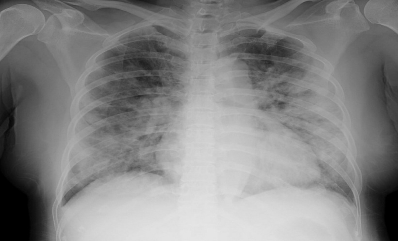
Mark van Buchem, Head of Radiology, Leiden University Medical Centre, the Netherlands, and Philippe Douek, Hospices Civils de Lyon, France, discussed their recent innovations developed in partnership with Philips. Van Buchem outlined the issues associated with MRI; its high run-time and resultant high cost coupled with poor patient experience. Together with Philips, van Buchem developed a tool named SmartSpeed. By under-sampling the K space during scanning in combination with prior knowledge and AI, they were able to decrease scan times eight-fold whilst increasing diagnostic accuracy. Douek worked with Philips during the COVID-19 pandemic to develop a quantitative assessment of long disease in patients with COVID-19. AI algorithms were able to quantify long lesions and fibrosis. Douek praised the work done during the pandemic, which improved accuracy of both diagnosis and prediction of ICU admission and death with just a single CAT scan.