This year, the European Congress of Radiology (ECR) took place online for the first time as a result of the ongoing coronavirus disease (COVID-19) pandemic. Over the 5-day scientific programme, there were 50 streamed sessions and over 1,800 abstract and poster presentations, with more than 200 virtual booths available to visit at the event. During the opening ceremony, Prof Boris Brkljačić, Chairman of the European Society of Radiology (ESR), announced that due to not having the usual time constraints of a physical congress, ECR 2020 will continue from July through to the end of December in the form of Highlight Weeks, each dedicated to a certain subspecialty or theme. On the decision to hold ECR 2020 online, Prof Brkljačić said: “Adaptation and innovation have always been strengths of the ESR, and having already established itself as a leader in online education over the past few years, the society rose to the challenge and made the decision to transform ECR 2020 into a fully online congress.”
The opening ceremony, like the entirety of the congress, captured the atmosphere of an onsite meeting. Prof Brkljačić welcomed attendees and thanked all radiographers and radiologists on the frontline of the pandemic, adding: “Your dedication for caring for patients, even when putting yourself at risk, is the strongest example of the compassion, understanding, and love that lies at the core of our professions.” Contributing to the progression of knowledge on COVID-19, the ESR have continually released educational resources and dedicated four sessions at ECR to the disease.
The theme of connecting science with art was present throughout the opening ceremony, with Prof Brkljačić drawing comparisons between the congress and Adagio in G Minor by Tomaso Albinoni, arguably one of the most famous pieces of classical music: “The spirit of this beautiful work is really in its sense of calm. This is the essence of Adagio, and that is our guiding philosophy for this year’s congress. To take a step back and think differently.”
Award winners were then announced, each preceded by their choice of music with a special significance to them, in a rendition by a four-piece band. ESR Honorary Membership was awarded to Prof Danta R. Casale Menier, Prof Yi-Hong Chou, and Prof Valerie P. Jackson, in recognition of their scientific excellence, international reputation, and achievements in national or international organisations. This year, Prof Richard FitzGerald, Prof Jim A. Reekers, and Prof Katrine Riklund received Gold Medals as acknowledgement for being outstanding scientists in the field of radiology or an allied science.
The plenary sessions were delivered by Prof Marie-Pierre Revel, in which the lessons and questions from the COVID-19 pandemic were considered; Prof Valérie Vilgrain, who discussed the resilience of the liver; Dr Bernd Montag, in which the movement towards digitalised healthcare was presented; and Prof Nenad Šestan, who detailed the formation of neural circuits.
Daily Table Talks took place throughout the congress, which were hosted by Prof Brkljačić and ESR Past President Prof Lorenzo Derchi. In these sessions, special guests were invited to discuss the scientific programme and current challenges in radiology, including clinical audits, lung cancer screening, and the special theme of Children in Focus.
This one-of-a-kind congress was truly spectacular, and, to quote Prof Brkljačić, “can only be described as a congress of the future.” We look forward to being able to attend what will surely be another great meeting next year in Vienna, Austria; until then, please enjoy the following review of the ECR 2020 Online Meeting.
Tin Filtration Reduces CT Dose and Improves Sacroiliitis Diagnosis
TIN FILTRATION can dramatically reduce radiation dose and therefore radiation exposure, which is in line with the ‘as low as reasonably achievable’ (ALARA) principle. One possible application of this technique is for confirmation of sacroiliitis diagnosis. In a study investigating tin filtrated ultra-low-dose CT (TFULDCT) in examining sacroiliac joints (SIJ) for sacroiliitis, it was shown that TFULDCT can more accurately identify whether inflammatory rheumatoid disease is present in SIJ joints compared with X-ray. Results from this study were presented at ECR 2020, and in an ECR Today news story.
CT is commonly used in diagnostic algorithms, and adherence to the ALARA principle is often achieved by restricting the extent of exposure, adapting scanning parameters, and adjusting images post-process. Tin filtration is another approach that can be taken; this reduces radiation dose by filtering low-energy photons, which are responsible for image contrast and noise reduction, between the X-ray generator and patient. The resultant reduction in radiation dose comes with the disadvantage of reduced image contrast and increased noise, limiting the use of tin filtration to distinguishing structures with significantly different densities.
In the pilot study, effective radiation doses and diagnostic usefulness of TFULDCT (median dose: 0.11 mSv) and X-ray (median dose: 0.25 mSv) were evaluated. The TSULDCT and X-ray images were independently evaluated by three radiologists, and findings were decided to be either clearly positive, clearly negative, or uncertain for the diagnosis of sacroiliitis. The results showed that TRULDCT had a significantly better overall diagnostic benefit than X-ray (80% versus 30%, respectively).
Lead author Dr Eva Korčáková, University Hospital in Pilsen, Pilsen, Czech Republic, commented: “Our results confirm that TFULDCT is able to more accurately decide the presence or absence of inflammatory rheumatoid disease of SIJ compared to X-ray.” She added that: “TFULDCT provides a convenient alternative where CT sensitivity is maintained but the dose decreases below the level of X-ray exposure.”
Evaluation of Bone Involvement in Stage IV Neuroblastoma with Diffusion-Weighted MRI
MANAGEMENT of patients with Stage IV neuroblastoma (NBL) requires accurate evaluation of metastatic bone disease including assessment of response to chemotherapy. Diagnosis with radiolabelled metaiodobenzylguanidine (MIBG) imaging is currently the gold standard; however, according to new research presented at ECR 2020, active metastatic bone lesions that are depicted on MIBG imaging are also evident on diffusion-weighted (DW) MRI, previously considered a limitation of MRI.
Here, researchers at SJD Barcelona Children’s Hospital, Barcelona, Spain, hypothesised whether MRI could detect bone involvement in NBL and if quantitative DW-MRI imaging is able to differentiate non-viable lesions from viable tumours.
In the retrospective study, 18 patients were recruited to compare whole-body DW-MRI and MIBG to assess the presence of metastatic osseous lesions in patients with Stage IV NBL who had received conventional chemotherapy. Results demonstrated a total of 171 lesions on all 18 patients. On MIBG, 73 bone lesions were reported as positive and nine MIBG bone lesions were not represented on DW-MRI. Coincidence between DW-MRI and MIBG was observed in 62 lesions and a statistically significant correlation was established between MIBG-positive bone lesions and a lower apparent diffusion coefficient value on DW-MRI.
Th evaluation of bone involvement in paediatric NBL has been considered a limitation of MRI because of its inability to differentiate between viable tumour and nonviable residual lesions. This is the first study comparing MIBG and DW-MRI in the evaluation of bone metastatic disease in paediatric NBL and results presented here suggest that active metastatic bone lesions that are depicted on MIBG imaging are also evident on DW-MRI and show restricted diffusion compared to ‘residual’ bone lesions that are not evident on MIBG.
While MIGB is the gold standard for the diagnosis of NBL, with a sensitivity and specificity of 90% and 100%, respectively, DW-MRI evaluation of bone involvement in Stage IV NBL is a valuable option and complementary use of whole-body DWI and MIBG scintigraphy can increase diagnostic accuracy in the evaluation of bone involvement.
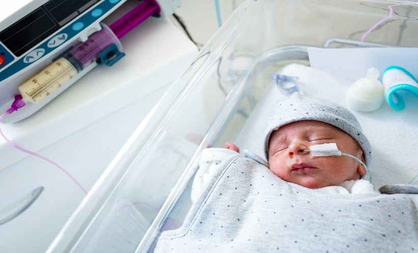
Predicting Fetal Lung Maturity with a Noninvasive Technique
NONINVASIVE sonographic techniques can be used to predict and evaluate fetal lung maturity. This is according to the results from a new study presented at ECR 2020, and in an ECR Today news story.
In the Minia University Maternity and Children’s Hospital in Minia, Egypt, 40 females between 28 and 40 weeks of gestation were enrolled in an observational prospective cohort study that used main pulmonary artery Doppler measurements to predict fetal lung maturity. In Egypt, approximately 7.3% of all live births are premature. Respiratory distress syndrome is a significant complication of prematurity and the most common cause of respiratory distress in premature infants, strongly associated with structural and functional immaturity of the lungs. This lack of full maturity in the lungs is the most common cause of death in neonates.
The risk of respiratory distress syndrome in infants can be predicted with the use of biochemical tests such as the lecithin/sphingomyelin ratio, which require invasive amniocentesis. Lung volume measurement, gestational age, epiphysis centres, placental grading, and estimated fetal weight are noninvasive methods that use ultrasound to evaluate fetal lung maturity but thus far, the techniques have not achieved success. The authors of the study aimed to explore alternative noninvasive ultrasound techniques for fetal lung maturity assessment using quantitative ultrasound texture analysis and pulmonary artery Doppler.
The main pulmonary artery Doppler diagnostic measurements included pulsatility index, resistance index, and acceleration time/ejection time, the accuracy for which was tested comparing clinical outcomes with the Doppler findings. Neonatal respiratory distress syndrome was diagnosed in nine fetuses (22%) and there was a significant correlation between low acceleration time/ejection time and respiratory distress syndrome development. The authors concluded that a noninvasive fetal lung maturity test and fetal pulmonary artery acceleration time/ejection time ratio are of similar effectiveness for predicting fetal lung maturity.
How to Manage Patient ‘No-Show’ in Radiology Departments
PREPROCEDURAL preparation is essential for nuclear medicine departments, and patient no-show, where patients do not turn up for their scheduled appointment, has a large impact on the productivity of hospitals. In an abstract presented at ECR 2020, results from a study in Doha showed the impacts no-show has on hospitals, and how to negate these.
Patient no-show is problematic for all hospital departments, and for radiology departments causes the issue of nonreusable radioactive material wastage, such as technetium 99m (Tc-99m), which ultimately leads to significant financial loss to the hospital.
In the study, between January and February 2019 loss to the hospital in terms of finance and time was calculated, based on the orders of Tc-99m and other radiopharmaceuticals and the length of appointments. A total of 84 out of 499 appointments were reported as no-shows during the 2-month study period. This 17% no-show rate caused a loss to the hospital of $39,000 USD and 864 hours wasted.
Reasons for no-show included illness, lack of transport, surgery, death in the family, or another appointment. The authors thereby decided to focus on patient education regarding the negative effects this has on the hospital, as well as an option to choose a preferred appointment date, rather than being assigned one by hospital staff. Additionally, the authors noticed that the no-show percentage reached as high as 22%, and suggested that a stricter no-show policy, such as a monetary deposit that is nonrefundable upon no-show, be implemented.
Following the distribution of educational leaflets, assignment of appointments according to patient preference, and hospital staff emphasis of the harmful impacts of missed appointments, the no-show percentage reduced to 13% (-4%).
The authors advocated for continuous training and education on the negative effects of patient no-show by hospital staff, improved communication skills training to receptionists, and use of emails as reminders. It is hoped that these changes will result in less financial loss to the hospital and shorter waiting times.
Angiography Assesses Effects of Decellularisation on Renal Transplants
FLUOROSCOPIC angiogram could be an important step in determining the transplant-readiness of decellularised kidneys. An Israeli study, presented at ECR 2020, has identified the value of the procedure to assess both vascular patency and perfusion following decellularisation.
Decellularised kidneys have had their cellular content removed, preserving the extracellular matrix and microstructures, biochemical composition, and cell-instructive signals, through a process of whole organ perfusion decellularisation. The intent of this technique is to remove immunogenic components so that transplant tissues can be available to any recipient, regardless of donor match. The technique relies on intact vasculature to circulate detergent-based solutions that dissolve cells and remove cellular antigens and DNA.
A study at a major transplant centre in Israel assessed the value of fluoroscopic angiography to reliably determine the integrity of arteries and veins of porcine kidneys, both pre- and postdecellularisation. Controlled flow conditions were used during fluoroscopic angiography to gauge vasculature patency and permeability via measurement of contrast media greyscale intensity and washout index.
The ex vivo angiograms revealed no difference in vessel patency following decellularisation, but did show significantly lower washout index in the porcine kidneys following the decellularisation process. The study identified delayed contrast clearance and increased vascular permeability, as well as evidence of extravasation of the contrast media into the kidney parenchyma. These changes were only evident in the kidneys following decellularisation.
The researchers undertaking the study, in a collaboration between the Department of Organ Transplant and Department of Radiology, Rabin Medical Center, Petah Tikva, Israel, highlighted the importance of these findings to the progress of developing bioengineered transplant organs: “Flow-controlled fluoroscopic angiography based on the proposed methodology is an accessible, accurate, and sensitive method that should be adopted as method-of-choice for evaluation of vascular integrity in bioengineered organs.”
Advancements in MRI Includes Grading Scale for Endolymphatic Hydrops
ENDOLYMPHATIC hydrops (EH) is the dilation of the endolymphatic section in the inner ear and compartments of the perilymphatic space. It occurs in inner-ear diseases and is acknowledged as the anatomical and pathological indicator of Ménière’s disease, a chronic condition with hallmark fluctuating low-frequency hearing loss, vertigo, tinnitus, and blockage sensations in the ear.
Autopsy, rarely carried out for inner ear pathology, was previously the only known method for EH diagnosis; therefore, research into Ménière’s disease and EH has been limited. Authors of a novel study from Pamplona, Spain, used information from patients with unilateral definite Ménière’s disease and aimed to evaluate the degree of interobserver similarities in both the detection and grading of EH in the cochlea and vestibule. The results for this study were presented at ECR 2020 and shared in an ECR Today news story.
In the study, vestibular EH was evaluated in 75 patients with diagnosed unilateral definite Ménière’s disease using cisternography, T2-FLAIR, and REAL IR imaging. In the clinically affected ear, EH was identified 90.6% of the time, and only 17.3% in the silent side. Three different visual scales to evaluate vestibular EH were compared: 4 stage, none/mild/moderate/severe; 3 stage, none/moderate/severe; and 2 stage, none/present. The researchers found that a 4-stage vestibular EH grading system provided the best interobserver consistency.
Of late, MRI has advanced significantly and is able to provide assessment in vivo of EH. The testing methods have been thoroughly researched and are now being implemented in the clinical field and will soon be utilised within radiology hospital departments. Studies thus far have investigated the validation and evaluation of treatment response in patients with Ménière’s disease by looking into the correlation between findings of MRI EH and clinical and cochleovestibular tests. The different scales for EH grading used in these studies prevent effect reproduction and comparability between studies, highlighting the importance of this novel study in determining the relationship between data in clinical studies. With this information, future research could establish the recommended scale for EH scale and provide clarity on study differences.
Cost-Effective Strategies for Solitary Pulmonary Nodule Diagnosis
SOLITARY pulmonary nodule (SPN) diagnostic accuracy is important because if early-stage lung cancer is treated with curative intent, the 5-year survival rates can be up to 70%. The diagnostic challenge associated with this is that not all SPN are a result of lung cancer, and accurate characterisation of the SNP can be expensive and inaccessible. However, dynamic contrast-enhanced CT (DCE-CT) has shown to be accurate and cost-effective, as presented in an abstract at ECR 2020 and in an ECR Today news story.
Nodule size is associated with the risk of malignancy, and thus directs the use of current management strategies. No follow-up is required for nodules <5 mm, but PET/CT or biopsy are used for further investigation of nodules >8 mm. In addition to the expensive costs associated with PET/CT and biopsy, access to PET/CT can be limited as it is not uniformly available at all hospitals.
In the large, multicentre trial, 312 patients with an SNP sized 8–30 mm were recruited across 16 hospitals in the UK. The participants underwent both PET/CT and DCE-CT, and biopsy/surgical resection or stability over the 2-years follow-up imaging period were used to determine final diagnosis of the nodule. DCE-CT uses intravenous iodine-based contrast to quantify the degree of enhancement of the nodules, which represents the vascularity, a highly sensitive and moderately specific marker for SPN. However, the trial concluded that PET/CT is more accurate for SNP diagnosis, but DCE-CT is more cost effective.
When the investigators explored ‘willingness to pay’ per correctly treated malignancy, DCE-CT was the preferred strategy when below £9,000 (€10,700). However, a combination of DCE-CT and PET/CT became the most-likely cost-effective strategy (probability equal to one) when society’s willingness to pay to get one more correctly treated malignancy was increased to £16,000 (€19,000). Authors of the study Prof Fiona Gilbert and Dr Jonathan Weir-McCall from the University of Cambridge, Cambridge, UK, noted: “These findings have significant implications, especially in light of the introduction of CT-based lung cancer screening.”
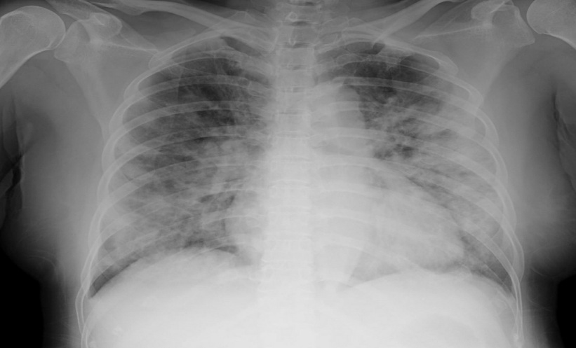
Radiation Dose Optimisation for Paediatric Chest Radiography
EXPOSURE to ionising radiation has been shown to generate a higher relative risk of leukaemia and brain, skin, thyroid, and breast cancer in children. Chest radiography is the most requested radiographical examination in paediatric patients (0–18 years old), and although it has a reasonably low exposure dose, a study presented at ECR 2020 sought to establish radiological exposure protocols for chest radiographs for this patient population, to reduce unnecessary exposure.
The study involved three steps: optimisation tests, quality evaluation, and clinical application. Variables that were accounted for included age, weight, height, projection of the exam, tube potential, exposure time, use of automatic exposure control, use of an anti-scatter grid, focus-detector distance, and kerma-area product. A sample of 44 examinations, 59.1% of which were male, were analysed, and the children were classified by weight.
Dose reductions dependant on weight class were applied using the protocol, which led to a 60% relevance for Class 2 (<5 kg), 75% for Class 3 (5–15 kg), 59% for Class 4 (30–50 kg), and 54% for Class 5 (50–80 kg). Statistical significance was reported both before and after optimisation and was significant for all weight classes.
Importantly, it was found that reducing exposure would not compromise the quality of the radiological examination. These results will guide clinicians in implementing an optimised cycle of the most frequently used radiographic examinations in paediatric patients, to minimise the harmful effects associated with diagnostic tests and therapeutic procedures that utilise ionising radiation.
Nanoparticles Make Microfractures Easily Visible on CT Images
MICROFRACTURES in bones can be difficult to detect due to the dense abundance of calcium in the bone, but if left untreated, they can develop into a complete bone fracture. In study results presented at ECR 2020 and summarised in an ECR Today news story, researchers showed that targeted nanoparticles could make microfractures profoundly visible.
Conventional CT cannot be used to identify microfractures due to the spatial resolution being approximately 0.500–0.625mm, and thus not allowing the detection of damage at the micrometre level; therefore, X-ray imaging-based techniques are normally used.
In the study, the researchers introduced colour to X-ray images using targeted nanoparticles comprising K-edge metals that are chemically ‘trained’ to locate microfractures, which then become visible with CT imaging. These particles differentially bind to the calcium in the microcracks, and, based on their distinctive K-edge energies, can be quantified using photon counting.
The K-edge nanoparticles accumulate in the microfractures and colourised voxels on the background of grey-scale X-ray are produced, allowing the identification of these contrast-enhanced targets. The researchers utilised MARS spectral CT technology equipped with Medipix detectors and MARS photon counting CT, and the resultant images discriminated microfractures from healthy bone with unparalleled clarity.
“Together with these clinically translatable ligand-directed particles, MARS can generate images that reveal abnormalities that usually remain undetected by conventional methods,” summarised Dr Dipanjan Pan, one of the researchers involved in the study, University of Illinois at Urbana-Champaign, Illinois, USA, and University of Maryland, Maryland, Baltimore, USA.
Intracranial Haemorrhage Detection by Artificial Intelligence
ARTIFICIAL intelligence (AI) supports on-call residents in detecting intracranial haemorrhage (ICH) at the emergency department of the Universitair Ziekenhuis Brussel (UZ Brussel), Brussels, Belgium. Here, the radiology workflow integrated an automated triage system which prioritises critical cases, based on the patient’s severity and urgency, ensuring that these patients are treated first. The data from this research were presented at ECR 2020 and summarised in an ECR Today news story.
To assess the performance of an AI tool using a deep learning algorithm for the detection of ICH on head CT exams with clinical workflow integration, researchers at UZ Brussel collected a dataset of 500 consecutive CT exams and their clinical reports between 1st September and 1st October 2019. CT exams were transferred for ICH detection by an AI tool, and the number of successfully processed studies by the AI were registered and the diagnostic performance, sensitivity, and specificity were calculated.
During the 1-month implementation phase, reports for 338 studies (77.6%) were generated by the algorithm and 22.4% failed, most likely because of technical issues. From these 388 studies, the AI tool labelled 31 (7.9%) as having ICH while the expert readers detected 37 (9.5%) with ICH. ICH detection between AI and expert readings was considered to be similar, and the algorithms performance revealed a 98% negative predictive value, 61% positive predictive value, and a sensitivity and specificity of 84%.
Dr Nina Watté, UZ Brussel, noted: “AI could automatically evaluate three-quarters of all head CT exams. With high specificity and negative predictive value, the AI tool shows the potential to rule out ICH, helping radiologists select which scans need to be viewed less urgently.” The potential for AI to support the selection of imaging modalities, diagnoses, and even in communication with colleagues and patients is vast. Looking forward, Dr Watté concluded: “This is an opportunity to leverage the power of AI to shape the radiologists of the future.”