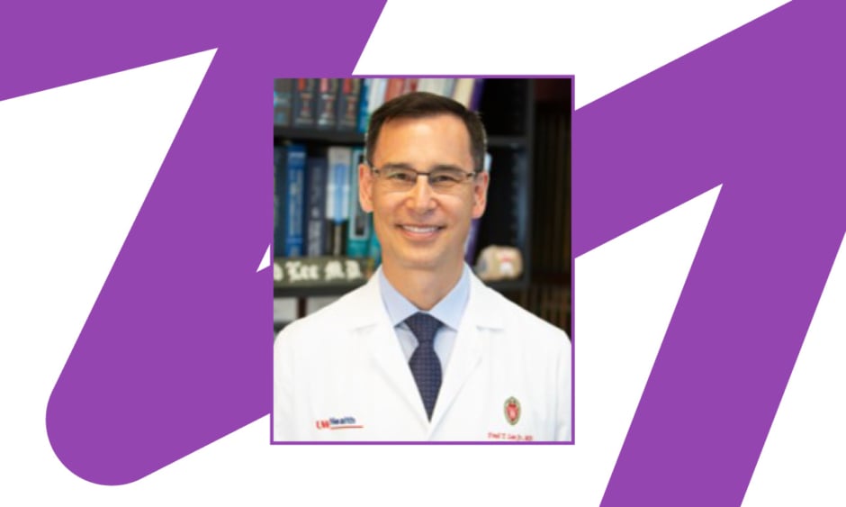
Fred T. Lee Jr. | Professor of Radiology, Biomedical Engineering, and Urology, the Robert A. Turrell Professor of Imaging Science; and the Chief of Abdominal Intervention at the Department of Radiology, University of Wisconsin School of Medicine and Public Health, Madison, Wisconsin, USA
Citation: EMJ Radiol. 2022;3[1]:34-38. DOI/10.33590/emjradiol/10199687. https://doi.org/10.33590/emjradiol/10199687.
![]()
With your experience in radiology and biomedical imaging, what initially sparked your interest in cancer imaging and interventions?
Having two parents who were diagnosed with cancer during my medical school years sparked my initial interest in the imaging of cancer. Our family had to live with the difficult trade-offs that all cancer patients need to make, and this got me thinking about less invasive and more effective methods to diagnose and treat cancer.
Your work to date has shifted to have a particular emphasis on percutaneous tumour ablation. As an early pioneer in this field, is there a particular event or person that encouraged you to go down this path?
My father was an early pioneer in the imaging and minimally invasive therapy of prostate cancer. He even diagnosed himself, and then decided to devote his remaining life to understanding and treating prostate cancer. In fact, for those of you out there who practise prostate MRI, he was the one that described the zonal anatomy of the prostate, was the first to use the prostate-specific antigen test in clinical practice, described prostate-specific antigen density, and was one of two people (Gary Onik being the other) to drive percutaneous cryoablation as a therapy for prostate cancer. After I joined the faculty at the University of Wisconsin, USA, in 1991, he would come to my lab, and we would perform animal studies together to try and understand ablation better. I knew that the prostate was not an organ that I wanted to spend a career working on, and so applied many of the lessons that he taught me to the liver, kidney, and lungs.
You initially studied at Boston University, Massachusetts, USA, then moved for your residency at the University of Rochester, New York, USA, and carried out your internship at the University of Massachusetts, USA. Where do you believe that you gained the most experience, and which had the largest impact on your subsequent career?
Looking back at my training, it was never about the ‘where’, but more about the ‘who’. I feel fortunate to have stumbled upon mentors that didn’t just teach me radiology, but rather how to approach problems, how to form and test a hypothesis, how to write, and, maybe most importantly, how to not give up in the face of seemingly insurmountable problems. For example, Jack Thornbury and Rick Katzberg (unfortunately both deceased) taught me the beauty of academic medicine, and Rob Lerner and Stan Weiss taught me the power of cross-sectional imaging, particularly ultrasound, as a method to place needles and devices. Another hugely motivating factor in my training was my residency classmates: Jim Bronson, Bevan Bastian, Jamey Schuster, and Dana Zacharewicz. I couldn’t have asked for better partners; we worked really hard together and encouraged each other. Medical training is tough, and if you don’t have great people around you sharing the journey, it’s easy to become discouraged
and cynical.
Do you think there are any misconceptions or challenges that the speciality of cancer imaging and intervention must overcome?
The entire concept of interventional oncology is fairly new, and I think we still struggle to be accepted on equal footing with medical, surgical, and radiation oncologists. I find this ironic because it is becoming increasingly obvious that image-guided therapies are here to stay, and will become more and more important to a wider segment of cancer patients as our techniques and devices become less invasive and more effective. As I look back at the last few decades, our progress is spectacular, particularly given the relatively short amount of time and small amount of funding we have received for our discoveries. For example, if one were to think about the total amount of dollars that have been spent on chemotherapies in the last two decades, if even a fraction of that amount had been applied to interventional oncology therapies, we would be in a very different place than we are today.
How do you believe your work as a founding member of the International Working Group for Tumour Ablation has impacted patients’ quality of treatment and quality of life?
The International Working Group was founded primarily by Nahum Goldberg, Damian Dupuy, and Luigi Solbiati in the early 1990s from a small group of us that were working in tumour ablation. We would meet at Gino’s Pizzeria during our time at the Radiological Society of North America (RSNA) and discuss every aspect of tumour ablation, including some of the hard things like complications and suboptimal results. In some ways, because there were so few of us performing tumour ablation at that time, it initially functioned as more of a support group than a scientific forum. Nahum was interested in finding a way to expand the impact of that initial small group, and so he started to champion tangible steps to push the field further along. For example, the original tumour ablation lexicon published in Radiology standardised how we talk about tumour ablation and was a by-product of the working group. Other important documents have since come out of the working group (or a subset of it) that have established standards and expectations for the practice of tumour ablation.
As an author whose work has resulted in over 200 scientific publications, where can we expect to see your focus lie in the coming years?
Over the past several years, I have mostly been working on histotripsy, a non-invasive, non-thermal, non-ionising method of tissue destruction that was invented at the University of Michigan, Ann Arbor, USA approximately a decade ago. The technology is very interesting to me because of the robotic control and automation used (we need more reproducibility and standardisation in interventional oncology), the high degree of precision, the ability to destroy virtually any size and shape of tumour, and the potential post-treatment immune effects. There’s a lot of work yet to be done, but the promise of better, faster, more automated, and less-invasive treatments make the effort worthwhile.
You have been featured in podcasts such as ‘The Kinked Wire’, where you spoke about the potential for histotripsy in cancer treatment. How do you think working in science communication helps to contribute to collective expertise and engagement?
In the early days of my career, science communication was simple: there were peer-reviewed journals, scientific meetings, and sometimes the print version of a specialty newspaper, such as this one. As they are highly curated and accurate, these modes of communication remain important, but they lack the ability to rapidly communicate with large numbers of physicians in a medium that they use daily. I personally don’t engage with social media, but can understand why some of my younger colleagues find it exceedingly useful and topical. Podcasts are one of my favourite methods to both communicate my own thoughts and hear about the work of others. There’s something about hearing the author explain their thought process in-depth that I find particularly valuable, and sometimes lacking from the more traditional modes of communication.
In 1995, when you established the tumour ablation laboratory at the University of Wisconsin, which was one of the first of its kind, what were the biggest challenges for the lab in achieving its initial goals?
Tumour ablation is particularly challenging to study in humans because we rely on imperfect imaging surrogates to determine the success or failure of treatment. In contrast, surgery has the advantage of a pathologic specimen to examine margins, microscopic pathology, genetic markers, etc. My initial purpose in establishing an animal laboratory to study ablation was to answer the simple but fundamental question: “How do you know you are doing what you think you are?” An animal lab therefore became a necessity in order to have access to tissue specimens after treatment and to correlate pathologic findings with imaging. It was a novel idea for its time, and grant funding was difficult because few reviewers had even heard of tumour ablation. For some reason, it seemed as though basic scientists with expertise in radiotherapy were the reviewers on all my early grant applications, and I wasted far too much effort explaining that interventional oncology treatments are indeed useful in patients. I am hoping that interventional oncology has now become so critical for modern cancer care that these arguments are a vestige of the past.
How have you acquired the leadership skills to establish and run your tumour ablation laboratory?
Leadership is all about finding the right people and letting them do their thing. If there’s anything that I’ve done right in my time at Wisconsin, it’s having the good fortune to work with great people who have carried our ideas forward.
What are some points of emphasis that you incorporate into practice to be the best interventional radiologist you can be?
One thing that I think is under-appreciated in the procedural specialties is the impact of a great team and environment around you. Too often, the physician tries to play too many roles when performing a complex procedure. In my opinion, the physician who is delivering the needle or catheter should be thinking only about getting the device into the right spot and not about sedation, monitoring the patient, running a machine, adjusting imaging etc. We have purposely set up our ablation service to reflect this strategy, and we have an entire anaesthesia team to perform jet ventilation, as well as nurses and technologists to position the patient and run the CT and ultrasound machines. Each person is highly experienced in their role, which allows the physician to concentrate on delivering the device to where it needs to go.
One other point of emphasis that we have incorporated into our practice is to track every important outcome of every procedure that we do, no matter how minor. We know our complication and failure rates in near real-time, and we can adjust on the fly if something systemic appears to be going wrong. Without tracking your procedural metrics, it’s very hard to improve.
Are there any innovations on the horizon, specifically in the field of radiology and cancer imaging and therapy, that you think are particularly noteworthy?
I’m particularly interested in the impact of robotics combined with artificial intelligence and machine learning on image-guided procedures. I believe that advances in these areas in the next decade will revolutionise how we do even the most mundane procedures. Along with making image-guided procedures less invasive and more effective, we need to automate, standardise, and measure what we do if we are going to improve our outcomes across populations.


