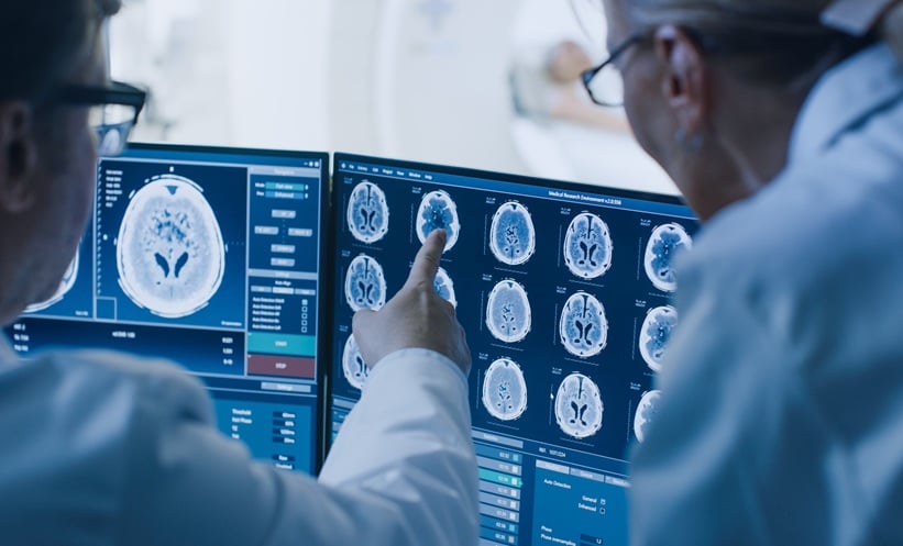
A NEW study has shown that combining neuroimaging with artificial intelligence (AI) can significantly enhance brain tumour diagnosis, achieving near-perfect accuracy in classifying tumour types.
Brain tumours remain among the most complex neurological conditions to diagnose, often requiring multiple imaging techniques and specialist interpretation. Magnetic resonance imaging (MRI) is widely used, but distinguishing between tumour types such as glioma, meningioma, and pituitary tumours can still be challenging.
As a result, there is growing interest in using machine learning to support clinical decision-making and improve diagnostic precision.
Brain Tumour Diagnosis Enhanced by AI
Researchers developed a fusion approach integrating MRI data with three machine learning models: convolutional neural networks (CNN), random forest (RF), and support vector machines (SVM). By combining structural imaging with advanced computational analysis, the system aimed to improve brain tumour diagnosis and classification accuracy.
The study analysed 7,023 MRI images spanning four categories: glioma, meningioma, pituitary tumour, and no tumour. Using a standard train–test split, all three models demonstrated exceptionally high performance. The CNN model achieved the highest accuracy at 99.29%, followed closely by RF at 99.06% and SVM at 98.36%.
These findings suggest that integrating multiple analytical approaches can capture subtle imaging features that may be missed by conventional assessment alone, strengthening brain tumour diagnosis in complex cases.
Why This Matters for Clinical Practice
Accurate classification is critical in brain tumour diagnosis, as treatment strategies and prognosis vary significantly between tumour types. Even small diagnostic errors can lead to inappropriate therapy or delayed intervention.
By improving classification accuracy, AI-driven tools could support radiologists and neurologists in making faster, more reliable decisions. This is particularly relevant in high-pressure clinical environments where rapid diagnosis is essential.
Limitations Highlight Need for Broader Validation
Despite the promising results, the authors highlighted key limitations.
The model was trained and tested on a single publicly available dataset, which may not reflect the diversity of real-world clinical populations.
Additionally, no external validation was performed, raising questions about generalisability.
Future Directions for Brain Tumour Diagnosis
The researchers suggested that future work should focus on multi-centre datasets, real-world clinical integration, and federated learning frameworks to enhance robustness.
Expanding these models across institutions could help ensure consistent performance in diverse healthcare settings.
If validated in clinical practice, this fusion approach could mark a significant step forward in brain tumour diagnosis, offering a powerful tool to support precision medicine in neurology.
Reference
Khan UA et al. Neuroimaging and machine learning fusion for improved brain tumor diagnosis and prognosis. Sci Rep. 2026;DOI:10.1038/s41598-026-50213-x.
Featured image: Gorodenkoff on Adobe Stock
- Author: