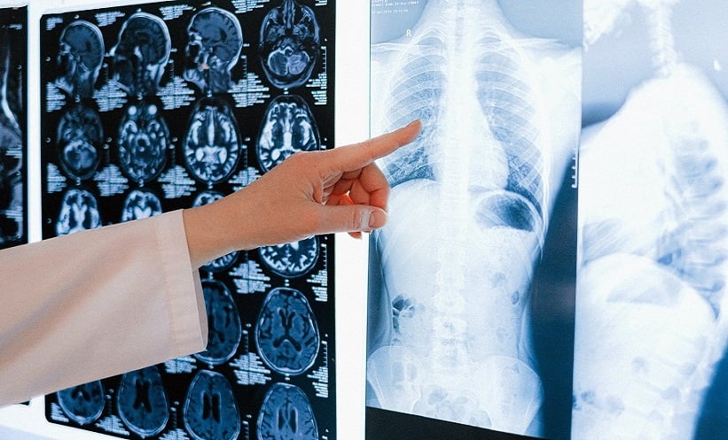
AN ARTIFICIAL intelligence (AI) system for humeral tumour detection has been developed, and can be applied to chest radiographs to evaluate impact on reader performance. This proposed AI tool has potential as a supportive diagnostic tool to alert radiologists about humeral abnormalities.
This study was of retrospective design, and included 14,709 chest radiographs between January 2000–December 2021. A total of 13,468 patients provided this dataset, of which there were proven normal (n=13,116) and humeral tumour (n=1,593) cases. A novel training method was introduced, called false-positive activation area reduction, to enhance the diagnostic performance by focusing on the humeral region. Receiver operating characteristic analyses were conducted for evaluation of model performance, studying the AI program and 10 radiologists.
False-positive activation area reduction application in the AI program improved performance compared with a conventional model, based on the area under the receiver operating characteristic curve (0.87 versus 0.82; P=0.04). The proposed AI system also demonstrated improved tumour localisation accuracy (80% versus 57%; P<0.001) and a false positive rate of 2%. The researchers were able to conclude that AI assistance improved radiologists’ sensitivity, specificity, and accuracy by 8.9%, 1.2%, and 3.5%, respectively (P<0.05 for all). This tool provided superior humeral detection on chest radiographs, and reduced false positives in tumour visualisation. Future study is expected to validate this practice on a wider scale.


