Key Summary:
- Four commercial AI tools did not improve chest X-ray diagnostic accuracy.
- AI reduced reading times by 6-17 seconds per case for some radiologists.
- Increased false positives highlight the potential risk of automation bias.

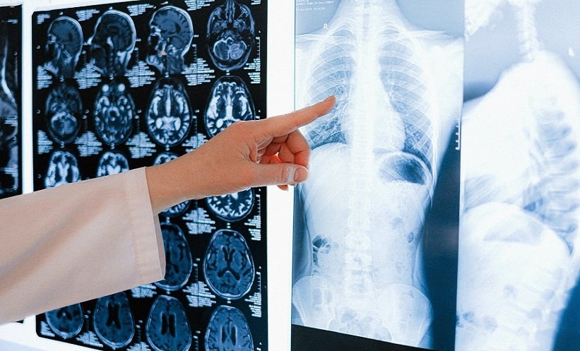
ARTIFICIAL intelligence (AI) may help radiologists interpret chest X-rays faster and with greater confidence, but this does not necessarily translate into greater diagnostic accuracy, according to new research.
A prospective study evaluating four commercially available AI systems found that AI assistance shortened interpretation times for some radiologists and increased confidence for most. However, none of the systems consistently improved diagnostic accuracy, and some increased false-positive findings.
Researchers at the Technical University of Munich, Germany, investigated how commercially available AI tools affected radiologists’ performance when interpreting chest radiographs.
The prospective crossover study included 1,200 consecutive patients undergoing chest radiography, comprising 1,861 radiographs. Five radiologists, with between 1 and 6 years of experience, assessed the images for pulmonary infiltrates, pleural effusions, mediastinal masses, pneumothorax, and pulmonary nodules.
Each radiologist interpreted the cases under five conditions: once without AI assistance and once with each of four different commercial AI algorithms. Sessions were separated by a 14-day washout period, while case order was randomised to reduce recall effects. Final clinical reports, supplemented by CT findings where available, served as the reference standard.
Alongside diagnostic performance, the researchers assessed interpretation time, diagnostic confidence, requests for senior review, and recommendations for additional CT imaging
Despite the additional support, AI did not improve overall diagnostic accuracy. For pleural effusions and pulmonary nodules, several combinations of radiologist and AI system actually reduced accuracy because of an increase in false-positive findings.
Nevertheless, the technology produced measurable workflow benefits. Three of the five radiologists interpreted images significantly faster with AI support, reducing median reading times by between 6 and 17 seconds per case. Four of the five readers also reported greater diagnostic confidence when using AI.
AI additionally reduced the need for senior consultation in selected reader-system combinations, while one combination resulted in fewer recommendations for further CT imaging.
The findings highlight an important distinction between improving radiology workflow and improving diagnostic performance. Increased confidence and faster reporting may benefit busy clinical environments, but could introduce risks if radiologists become more likely to accept incorrect AI-generated findings.
The researchers highlighted automation bias as a particular concern, suggesting that AI systems should be carefully adapted and evaluated within individual clinical settings rather than assumed to improve performance universally.
Further clinical trials will also be needed to determine whether workflow improvements associated with AI ultimately translate into better patient outcomes.
The findings suggest that the value of AI in chest radiography may lie not simply in whether algorithms can detect abnormalities, but in how effectively radiologists and AI systems can work together without compromising diagnostic accuracy.
Reference
Lemke T et al. Artificial Intelligence-Assisted Chest Radiography: A Prospective Crossover Multi-Reader Study on Diagnostic Performance and Workflow Efficiency. Academic Radiology. 2026. doi:10.1016/j.acra.2026.08.014.
Featured image: See Less on AdobeStock
Each article is made available under the terms of the Creative Commons Attribution-Non Commercial 4.0 License.