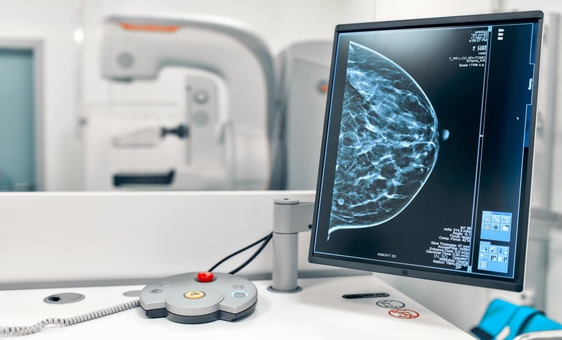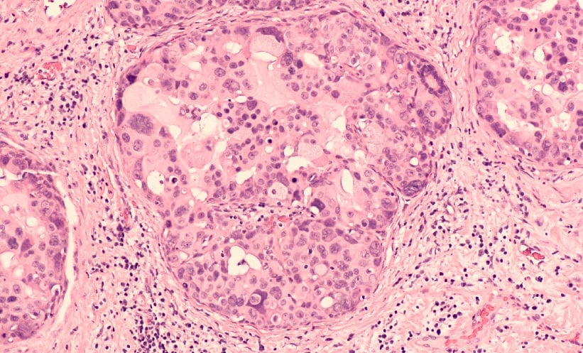
CONTRAST-ENHANCED mammography is more effective than low-energy image alone in diagnosing ductal carcinoma in situ (DCIS), a non-invasive form of breast cancer where abnormal cells remain confined within the milk ducts and have not invaded surrounding tissue. It also appears to improve assessment of lesion extent and recognise imaging differences across tumour grades.
Grade-Dependent Imaging Patterns in DCIS
Researchers conducted a single-centre retrospective analysis of 190 patients with pure DCIS who underwent contrast-enhanced mammography within two weeks prior to surgery. A total of 196 lesions were evaluated, including six patients with bilateral disease. Imaging was assessed to determine diagnostic performance across different pathological nuclear grades.
While contrast-enhanced mammography showed similarly high sensitivity across all nuclear grades, the study noted that DCIS subtypes displayed distinct imaging characteristics. High-grade DCIS was more likely to present with calcifications, larger lesion size, and greater enhancement conspicuity compared with lower-grade disease. In contrast, low-grade DCIS showed fewer pleomorphic and fine linear branching calcifications than higher-grade lesions.
However, enhancement patterns such as percentage uptake and morphology did not significantly differ between grading groups, suggesting some shared imaging behaviour despite histological variation.
Higher Detection Rates with Contrast-Enhanced Mammography
The study found that contrast-enhanced mammography demonstrated higher sensitivity for DCIS overall compared with low-energy imaging alone, with 89.3% versus 72.4%. This advantage was consistent across calcified and non-calcified lesions, as well as across low-, intermediate- and high-grade disease. Sensitivity was notably improved in non-calcified DCIS, where detection rose from 39.4% with low-energy imaging to 76.1% using contrast-enhanced mammography.
This is clinically relevant as non-calcified lesions are usually more challenging to identify on conventional mammographic imaging.
Implications for Breast Cancer Imaging Pathways
The results indicate that contrast-enhanced mammography was superior to low-energy imaging alone in demonstrating the full extent of DCIS lesions. This improved visualisation may be clinically relevant when planning surgical or therapeutic approaches, where accurate delineation of disease boundaries is important.
As a single-centre retrospective study, the findings may not be fully generalisable across broader populations or different imaging settings. Further research is needed to validate performance in multi-centre cohorts and assess how contrast-enhanced mammography integrates into routine diagnostic pathways.
Overall, the findings support contrast-enhanced mammography as a more sensitive tool for DCIS detection than low-energy imaging alone, with consistent performance across tumour grades. The ability to better define lesion extent and reveal grade-related imaging differences may help refine diagnostic confidence and improve preoperative assessment in clinical practice.
Reference
Wang L et al. Preoperative contrast enhanced mammography evaluation of breast ductal carcinoma in situ and comparative analysis with pathological grading. Clin Radiol. 2026;DOI:10.1016/j.crad.2026.107392.
Featured image: David A Litman on Adobe Stock
- Author: