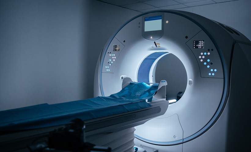
OVARIAN tumours classified as high risk on ultrasound could be further characterised using contrast-enhanced computed tomography (CE-CT), according to a retrospective study.
The findings suggest that CE-CT may help distinguish benign, borderline, and malignant epithelial ovarian tumours, supporting more individualised decisions about surgical management.
Assessing High-Risk Ovarian Tumours
The study included 210 women with 258 adnexal lesions who underwent preoperative transvaginal ultrasound and abdominopelvic CE-CT between September 2017 and December 2022. Researchers assessed lesion morphology and enhancement features to classify tumours as benign, borderline, or malignant.
Lesions classified as O-RADS 4 are considered intermediate risk, while O-RADS 5 indicates high risk. However, both categories can include borderline ovarian tumours (BOTs) and invasive epithelial ovarian tumours, which may require different surgical approaches.
CT Demonstrates Diagnostic Accuracy
CE-CT achieved a diagnostic accuracy of 88.8% across the three categories. When analysed according to ultrasound risk classification, accuracy was 87.5% among O-RADS 4 lesions and 90.0% among O-RADS 5 lesions.
In an exploratory analysis excluding patients with benign tumours, CE-CT also demonstrated greater accuracy than ultrasound in distinguishing BOTs from invasive carcinomas. Diagnostic accuracy was 89.3% with CE-CT, compared with 72.0% using O-RADS US categories alone.
However, some lesions were incorrectly classified. In particular, borderline tumours were sometimes interpreted as malignant, while certain benign lesions were also overestimated. Researchers noted that CE-CT enhancement patterns and cancer antigen-125 levels alone were not sufficient to reliably identify every tumour subtype.
Implications for Surgical Planning
The findings suggest that CE-CT could complement O-RADS US by providing additional information about primary tumour characteristics and disease extent. This may help clinicians better distinguish between patients with benign, borderline, and invasive tumours, supporting more individualised preoperative decision-making.
The study was limited by its retrospective, single-centre design, researchers highlighted the need for larger prospective studies to validate the findings.
Reference:
Yang S et al. Added Value of Contrast-Enhanced CT for Risk Stratification of Patients with Epithelial Ovarian Tumors Categorized as O-RADS US 4 and 5. J Am Coll Radiol.2026;23(11):2088-97.