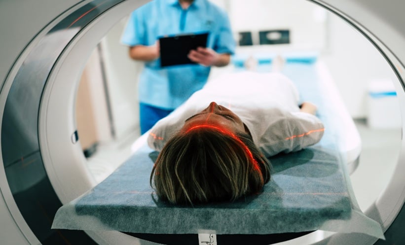
DISTINGUISHING between different conditions of the spine is important in order to provide patients with the correct diagnosis and treatment. Paediatric myelin oligodendrocyte glycoprotein demyelination (MOGAD), multiple sclerosis (MS), and seronegative monophasic demyelination can all affect the spine and have overlapping symptoms. Therefore, researchers must explore techniques that can help separate these conditions.
In a recent study, scientists used MRI to distinguish MOGAD from MS and seronegative monophasic demyelination. The researchers recruited 107 children to take part in this study, with an approximately equal split between young males and females. On average, the median age of disease onset was 11 years of age. Out of the large sample, 40 children had MOGAD, 21 had MS, and 46 had seronegative myelitis.
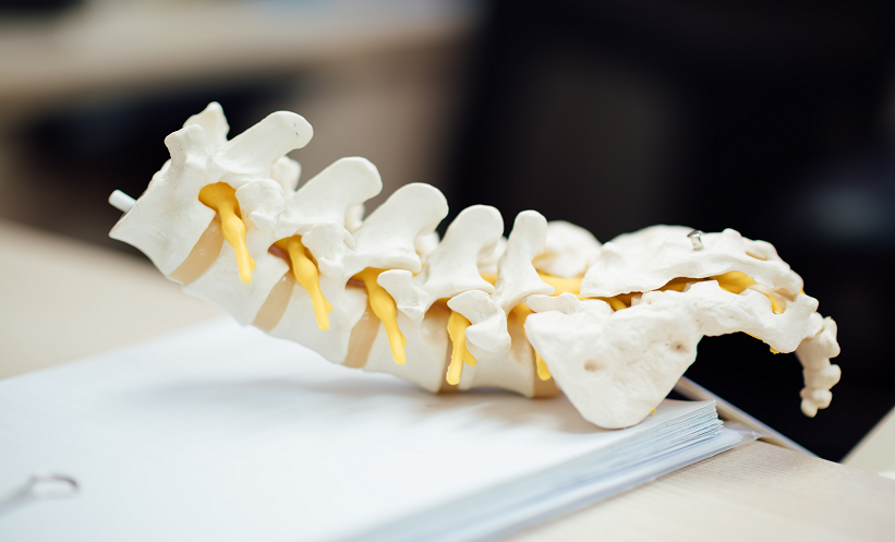
The involvement of the spinal cord was assessed in 324 MRI sequences, with the features and scores from this step reviewed with a 5-year follow-up period. The scientists discovered important features of MOGAD, notably enhancement of the leptomeningeal covering of the cord. Extensive spinal cord lesions were very common in patients with MOGAD and occurred in 75% of children. Interestingly, extensive lesions were less common in seronegative myelitis (43%) and rare in MS (5%).
Other important features included axial grey matter T2-hyperintensity and ‘snake-eye appearance’. The formerly mentioned characteristic was observed in 63% of patients with MOGAD and 33% of participants with seronegative myelitis. All these facets could help physicians identify MOGAD and encourage them to send serum for MOG-IgG testing.
Brenda Banwell, Children’s Hospital of Philadelphia, Perelman School of Medicine, University of Pennsylvania, USA, shared her concluding remarks: “These findings suggest that several features may help identify children at presentation who are more likely to have myelitis associated with MOGAD. Prominent involvement of grey matter and leptomeningeal enhancement are common in paediatric MOGAD, although the pathological underpinning of these observations requires further study.”