EPICARDIAL adipose tissue (EAT) volume, quantified on cardiac magnetic resonance imaging, may provide radiologists with a useful tool to identify patients at higher risk of heart failure with preserved ejection fraction (HFpEF) and its complications.
HFpEF is characterised by impaired ventricular filling despite a normal ejection fraction, contributing to a growing share of heart failure cases.
The findings position cardiac magnetic resonance imaging derived EAT volume as a potential imaging biomarker that reflects both disease severity and prognosis, expanding the role of cardiac MRI beyond conventional functional assessment.
Imaging Biomarker Gains Traction HFpEF
With HFpEF prevalence increasing and diagnostic pathways often complex, there is rising interest in imaging markers that can refine risk stratification.
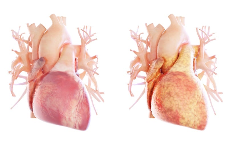
Epicardial adipose tissue, located between the myocardium and visceral pericardium, has emerged as a candidate due to its proximity to cardiac structures and potential pathophysiological relevance.
Cardiac Magnetic Resonance Shows Clear Risk Gradient
In this retrospective analysis, investigators used cardiac magnetic resonance imaging to assess 117 patients with HFpEF, 62 high-risk individuals, and 65 healthy controls.
EAT volume and left ventricular strain parameters were quantified using CVI42 software, reflecting routine post-processing workflows in cardiac imaging.
The EAT index demonstrated a stepwise increase across groups, from 20.32 ml/m2 in controls to 31.98 ml/m2 in high-risk individuals and 48.21 ml/m2 in HFpEF patients. EATi also showed good discriminatory performance, separating controls from high-risk individuals and distinguishing high-risk patients from those with established HFpEF.
Multivariable modelling incorporating strain parameters identified EATi and left ventricular global longitudinal strain as independent indicators linked to HFpEF, reinforcing the complementary value of structural and functional imaging markers.
Prognostic Value Extends Beyond Diagnosis
Follow-up data from 228 participants over a median of 31 months showed that higher EAT index was linked to an increased risk of heart failure readmission or all-cause mortality. Incremental rises in EAT index were also associated with higher risk of heart failure readmission or all-cause death, suggesting a role in prognostic assessment by enabling the identification of high-risk patients.
Implications for Radiology Practice
These findings support the integration of EAT volume assessment into routine cardiac magnetic resonance imaging interpretation, particularly in patients with suspected or established HFpEF. The ability to extract incremental diagnostic and prognostic information from standard imaging datasets may enhance multidisciplinary decision-making.
However, the retrospective design and need for prospective validation remain important limitations. Further research will be needed to determine how best to incorporate EAT quantification into clinical workflows and whether it can guide management strategies.
Reference
Zhang Y et al. Analysis of cardiac magnetic resonance characteristics in patients with ejection fraction preserved heart failure. Clin Radiol. 2026;DOI: 10.1016/j.crad.2026.107348.
Featured image: Sebastian Kaulitzki on Adobe stock
- Author: