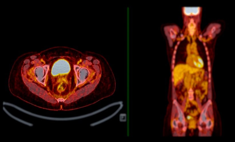
AS the use of antiamyloid therapies expands, amyloid PET is expected to play an increasingly central role in the diagnosis and management of patients with cognitive impairment. While previous research has often relied on standardised image acquisition and quantitative interpretation, real-world clinical settings typically depend on local, visually based reads, raising concerns about the consistency and accuracy of these assessments. A key finding from a new US-based study shows that local visual interpretation of amyloid-PET scans agrees with quantitative reads 86.3% of the time.
This cross-sectional quality improvement study analysed data from the Imaging Dementia-Evidence for Amyloid Scanning (IDEAS) initiative, involving 294 imaging facilities across the United States. Researchers included 10,361 Medicare patients aged 65 or older who had cognitive decline where Alzheimer’s disease was under diagnostic consideration. Amyloid-PET scans using [18F]florbetapir, [18F]florbetaben, or [18F]flutemetamol were interpreted visually by site radiologists or nuclear medicine specialists following approved guidelines. These were compared with quantitative scan assessments using the Centiloid (CL) scale, applying a previously validated autopsy-based threshold of 24.4 CL for scan positivity.
Of the 10,361 analysed scans, 6,332 (61%) were read as positive visually and 6,121 (59%) were positive quantitatively. Overall agreement between visual and quantitative classifications reached 86.3% (95% CI: 85.7%–87.0%), with a Cohen’s κ of 0.72. Concordance was higher among females (87.4% vs. 85.2% in males; p=0.001), White patients (86.6% vs. 84.5% in non-White; p=0.046), and when using [18F]flutemetamol or [18F]florbetaben compared with [18F]florbetapir (p<0.001). Discordance was more common in borderline CL values (10–40 CL), accounting for most mismatches.
The study confirms that visual reads, when performed by trained clinicians, provide highly reliable results that align well with quantitative measures, offering reassurance for everyday clinical use of amyloid-PET scans. Nonetheless, limitations include the cross-sectional design, reliance on a single quantitative threshold, and underrepresentation of certain racial and ethnic groups. Given the increasing clinical demand for amyloid imaging, these findings support the practical utility of visual PET interpretation while highlighting areas such as borderline cases, where additional quantification may still add value.
Reference
Zeltzer E et al. Concordance Between Amyloid-PET Quantification and Real-World Visual Reads. JAMA Neurol. 2025;DOI: 10.1001/jamaneurol.2025.2218.