BACKGROUND AND AIMS
Fragmentation of in vitro fertilisation (IVF) embryos is used as a marker for embryo deselection.1 Extensive fragmentation may be associated with reduced blastocyst formation and increased incidence of chromosomal abnormalities.2 However, some of those embryos may still lead to pregnancy and delivery of a healthy child. Success rate prediction in these embryos is still an open question. Moreover, the cut-off of fragmentation rate that still enables achievement of pregnancy is not clearly defined.3 It was hypothesised that using general and lab-specific models based on time-lapse technology may contribute to the selection of embryos with fragments.
MATERIALS AND METHODS
In this retrospective study, 4,210 embryos were analysed. They were incubated to the blastocyst stage in an EmbryoScope™ (Vitrolife, Gothenberg, Sweden) between 2013 and 2019 and included 379 embryos with >5% fragmentation. Embryos were selected for transfer or freezing versus deselection, based primarily on the general model for Day 5 development, provided by Vitrolife, and then re-examined by the laboratory’s specific algorithms.
Participants, Materials, Setting, and Methods
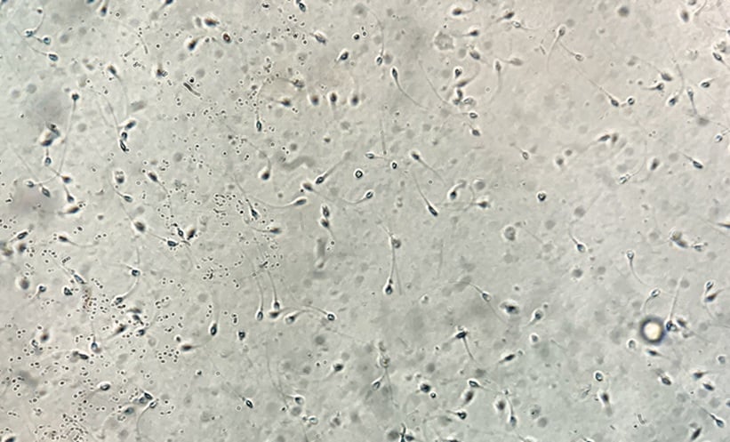
Embryo fragmentations were measured using EmbryoScope tools by a senior embryologist. Percentage of fragmentation was documented twice for every embryo: at the first cell division and at their maximum volume. The patterns of fragment accumulation during embryo development were followed. Data were analysed using statistical methods for fragmentation with regards to patient age, insemination method, blastocyst formation, embryo transfer or freezing, known implantation data of the embryo, clinical pregnancy, and live birth rate.
RESULTS
The specific model score and a fragmentation percentage of up to 32% were found to be independent variables. Embryos with up to 20±12% fragmentation still had a high score according to the first division time and were usually transferred or cryopreserved. Significant differences in the fragmentation rate were found between embryos which reached the blastocyst stage and embryos which failed to develop (15±11% and 36±20%, respectively; p<0.0001).
Fragmentation usually appeared at the first division and this worsened during the third or fourth divisions. While no difference was found in fragmentation between embryos of standard IVF or intracytoplasmic sperm injection, age had a significant negative effect on fragmentation (p<0.0001). In this population of fragmented embryos, 33 out of 379 embryos resulted in the delivery of a healthy child, 104 failed to develop, and information was not available for the remaining 242, mainly because they were frozen without thawing.
In 92.8% of patients, more than one embryo had fragments. In 64% of patients, fragmented embryos were found in more than one cycle. All positive known implantation data had a maximum of 30% fragmentation, except for one embryo with 43% fragmentation that was successfully implanted, and a healthy baby was born.