Deep Learning-Based Quantification of Traction Bronchiectasis Severity For Predicting Outcome in Idiopathic Pulmonary Fibrosis - European Medical Journal

Deep Learning-Based Quantification of Traction Bronchiectasis Severity For Predicting Outcome in Idiopathic Pulmonary Fibrosis

2 Mins
Respiratory
Authors:
*F. Felder,1 Y. Nan,1 G. Yang,1 J. Mackintosh,2 L. Calandriello,3,4 N. Goh,5-7 P. Hopkins,2,8 Y. Moodley,9 P. Reynolds,10 T. Corte,11 V. Navaratnam,12,,13 S. Walsh1
  • 1. National Heart and Lung Institute, Imperial College London, UK
  • 2. Queensland Lung Transplant Service, The Prince Charles Hospital, Brisbane, Australia
  • 3. Department of Diagnostic Imaging, Radiotherapy, Oncology, and Hematology, Agostino Gemelli University Policlinic, Rome, Italy
  • 4. Istituto di Ricovero e Cura a Carattere Scientifico (IRCSS), Rome, Italy
  • 5. Department of Respiratory and Sleep Medicine, Austin Health, Melbourne, Australia
  • 6. Institute for Breathing and Sleep, Melbourne, Australia
  • 7. University of Melbourne, Australia
  • 8. Faculty of Medicine, University of Queensland, Brisbane, Australia
  • 9. School of Medicine and Pharmacology, University of Western Australia, Perth, Australia
  • 10. Royal Adelaide Hospital Chest Clinic, Adelaide, Australia
  • 11. Respiratory Medicine, Royal Prince Alfred Hospital, Sydney, Australia
  • 12. Department of Respiratory Medicine, Sir Charles Gardiner Hospital, Perth, Western Australia, Australia
  • 13. University of Western Australia Medical School, Western Australia, Australia
*Correspondence to [email protected]
Disclosure:

Please click here for author disclosures.

Acknowledgements:

The authors would like to acknowledge and thank Mario Silva, University of Parma, Italy; Ian Glaspole, Alfred Hospital, Melbourne, Australia; Wendy Cooper, New South Wales Health Pathology, Sydney, Australia, Royal Prince Alfred Hospital, Sydney, Australia, and University of Sydney, Australia; Christopher Grainge, John Hunter Hospital, Australia; and Athol Wells, National Heart and Lung Institute, Imperial College London, UK, and Royal Brompton Hospital, London, UK, for their contributions to this research, and co-authorship of the original abstract presented at the European Respiratory Society International Congress 2023.

Citation:
EMJ Respir. ;11[1]:76-78. DOI/10.33590/emjrespir/10307156. https://doi.org/10.33590/emjrespir/10307156.
Keywords:
Artificial intelligence, high-resolution CT (HRCT), interstitial lung disease, predictor of mortality, predictor of progression, prognosis, pulmonary fibrosis, traction bronchiectasis.

Each article is made available under the terms of the Creative Commons Attribution-Non Commercial 4.0 License.

BACKGROUND AND AIMS

Idiopathic pulmonary fibrosis (IPF) is inexorably progressive. Like IPF, other fibrotic lung diseases can exhibit this progression, and these are called progressive fibrotic phenotypes. Early identification of these phenotypes is crucial to initiate treatment promptly.1-4

The severity of traction bronchiectasis, when visually assessed, is a strong predictor of mortality in patients with fibrotic lung disease. However, semi-quantitative methods have limitations, which can be addressed by the application of quantitative methods.5-7

The objective of this investigation is to explore the prognostic utility of a novel deep-learning algorithm in quantifying the severity of traction bronchiectasis in patients with IPF, who are enrolled in the Australian IPF Registry (AIPFR).

MATERIALS AND METHODS

Visual evaluation of high-resolution CT (HRCT) from the AIPFR was performed by two expert thoracic radiologists evaluated. Total airway volume (TAV) was quantified using a novel 3D U-Net-based deep-learning algorithm. Systematic objective fibrotic imaging analysis algorithm (SOFIA) usual interstitial pneumonia (UIP) probability scores were obtained using a previously reported deep-learning algorithm, trained in the identification of UIP features.5 The prognostic utility of automated airway volume quantification was evaluated against conventional measures of disease severity and SOFIA-based UIP probability scores.

RESULTS AND DISCUSSION

In total, 217 HRCTs were amenable to algorithmic airway volume quantification. TAV was an independent predictor of mortality when controlling for visual-based evaluation of total fibrosis extent (hazard ratio [HR]: 1.96; p<0.0001), % predicted forced vital capacity (FVC; HR: 2.15; p<0.0001), or the composite physiologic index (CPI; HR: 1.52; p=0.02). On bivariable analysis, both TAV (HR: 2.13; p<0.0001) and SOFIA-UIP probability (HR: 1.30; p<0.0001) independently predicted mortality. Patients were divided based on INBUILD criteria (Group 1: ‘UIP-like fibrotic pattern’, HRCT showing UIP or probable UIP, or surgical lung biopsy showing possible, probable, or definite UIP; and Group 2: ‘other fibrotic patterns’, remaining patients).1 On bivariable analysis with total fibrosis extent, TAV independently predicted mortality in Group 1 (HR: 1.50; p=0.03), and was the only predictor of mortality (HR: 5.33; p<0.0001) in Group 2. An increase in TAV of 1% of total lung volume was associated with a three-fold increased likelihood of developing progressive disease (FVC/diffusing capacity of the lungs for carbon monoxide decline of 10% and 15%, respectively, at 12 months; odds ratio: 3.04; p=0.009) when controlling for total fibrosis extent.

IPF exhibits an unpredictable and progressive clinical course. This means that, despite treatment, IPF is inherently progressive, akin to other interstitial lung diseases categorised as progressive phenotypes. Recognising progression is crucial to avoid delaying the onset of treatment and preventing clinical deterioration, ensuring timely intervention for the disease.

For IPF, progression is typically defined as a 10% or greater decline in FVC, or a 15% or greater decline in diffusing capacity of the lungs for carbon monoxide over 12 months, or sustained over 18 months, or in cases of death or transplantation.8,9

FVC is the most widely accepted marker and primary endpoint in clinical trials. However, it has limitations, like data loss, variability, and susceptibility to the influence of emphysema, prompting the exploration of alternative markers.

CT holds promise as a prognostic marker. Traction bronchiectasis has proven to be a powerful sign of progression. Yet, most studies evaluating them rely on semi-quantitative methods, which are susceptible to low reproducibility and inter-observer variability, and are unable to capture subtle changes over a short timeframe.5-7,10,11

TAV objectively identifies patients at risk of progression, aiding clinical decision-making and stratification of patients in clinical trials. Compared to FVC, TAV is a reliable short-term evaluation method (Figure 1).

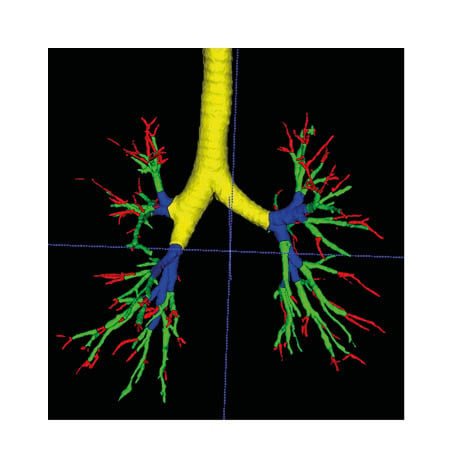
Figure 1: Segmentation of the airway.
The terminal bronchus is shown in red, the small bronchus in green, the medium bronchi in blue, and the large bronchi in yellow.

CONCLUSION

In IPF, automated quantification of TAV predicts mortality independently of total fibrosis extent on HRCT, and can be used to identify patients at risk of progression at 12 months.

References
Flaherty KR et al.; INBUILD Trial Investigators. Nintedanib in progressive fibrosing interstitial lung diseases. New Engl J Med. 2019;381(18):1718-27. Walsh SLF et al. Deep learning-based outcome prediction in progressive fibrotic lung disease using high-resolution computed tomography. Am J Respir Crit Care Med. 2022;206(7):883-91. Nasser M et al. Progressive fibrosing interstitial lung disease: a clinical cohort (the PROGRESS study). Eur Respir J. 2021;57(2):2002718. Takei R et al. Prevalence and prognosis of chronic fibrosing interstitial lung diseases with a progressive phenotype. Respirology. 2022;27(5):333-40. Walsh SLF et al. Deep learning for classifying fibrotic lung disease on high-resolution computed tomography: a case-cohort study. Lancet Respir Med. 2018;6(11):837-45. Jacob J et al. Serial CT analysis in idiopathic pulmonary fibrosis: comparison of visual features that determine patient outcome. Thorax. 2020;75(8):648-54. Walsh SLF et al. Imaging research in fibrotic lung disease; applying deep learning to unsolved problems. Lancet Respir Med. 2020;8(11):1144-53. Jo HE et al. Baseline characteristics of idiopathic pulmonary fibrosis: analysis from the Australian Idiopathic Pulmonary Fibrosis Registry. Eur Respir J. 2017;49(2):1601592. Erratum in: Eur Respir J. 2017;49(3):1651592. Jo HE et al. Disease progression in idiopathic pulmonary fibrosis with mild physiological impairment: analysis from the Australian IPF registry. BMC Pulm Med. 2018;18(1):19. De Giacomi F et al. Short-term automated quantification of radiologic changes in the characterization of idiopathic pulmonary fibrosis versus nonspecific interstitial pneumonia and prediction of long-term survival. J Thorac Imaging. 2018;33(2):124-31. Hwang J-H et al. Longitudinal follow-up of fibrosing interstitial pneumonia: relationship between physiologic testing, computed tomography changes, and survival rate. J Thorac Imaging. 2011;26(3):209-17.

Please rate the quality of this content

As you found this content interesting...

Follow us on social media!

We are sorry that this content was not interesting for you!

Let us improve this content!

Tell us how we can improve this content?