Abstract
Orthotropic liver transplantation poses an anaesthesiological challenge in patients who are cirrhotic and complexed by hepato-pulmonary syndrome (HPS) and porto-pulmonary hypertension (PoPH). This case report features the presence of two opposing pulmonary manifestations of advanced cirrhosis in a single patient and delineates the nuances in diagnosis and management for both HPS and PoPH. It then discusses the anaesthesia considerations of liver transplantation for this subset of patients. A female in her 60s with portal hypertension secondary to primary biliary cholangitis and autoimmune cirrhosis developed hypoxaemia refractory to oxygen supplementation in the setting of vasodilatory treatment for moderate PoPH. Progression of the principle cirrhosis several years after a transjugular intrahepatic portosystemic shunt procedure lead to the conversion of disease pathophysiology to hepatopulmonary syndrome via pulmonary arteriovenous malformation formation. Intrapulmonary shunting was detected through echocardiographic microbubble study, liver transplantation was the most evidence-based treatment route for this transformed HPS. It is therefore of clinical importance to distinguish between HPS and PoPH in patients with cirrhosis presenting with positional dyspnoea, because these two are similar but antagonistic disease processes requiring distinct treatment management with differing indications and anaesthesia considerations as it pertains to liver transplantation.
Key Points
1. Hepato-pulmonary syndrome (HPS) causing vascular dilatation and porto-pulmonary hypertension (PoPH) causing vascular constriction represent two of the three pulmonary manifestations of decompensated liver cirrhosis and can have temporal roles in a singular patient.2. While liver transplantation is the definitive therapy for HPS, elevated pulmonary arterial pressures exceeding 50 mmHg in severe PoPH contraindicate liver transplantation prior to pulmonary arterial hypertension therapy in patients dealing with both HPS and PoPH.
3. In the treatment of PoPH, titration of pulmonary vasodilators is necessary prior to liver transplantation, with therapies continued uninterrupted peri- and intra-procedurally.
BACKGROUND
With a prevalence of 4–47% in patients who are cirrhotic, hepatopulmonary syndrome (HPS) is just one of three pulmonary manifestations of chronic liver disease alongside porto-pulmonary hypertension (PoPH), which causes pulmonary vascular constriction, and hepatic hydrothorax, which causes translocation of ascitic fluid secondary to diaphragmatic defects.1 According to the European Respiratory Society (ERS) Task Force in 2004, HPS is characterised by a triad of: a) liver disease and/or portal hypertension; b) partial pressure of O2 <80 mmHg or Alveolar-arterial gradient >15–20 mmHg for patients under and over the age of 65 years, respectively; and c) intrapulmonary vascular dilatation visualised by contrast-enhanced microbubble echocardiogram or radio-labelled albumin perfusion scanning.2 The four-level severity of HPS is staged by arterial O2 capacity determined by arterial blood gas analysis, and ranges from mild, with pulmonary pressure of O2 in arterial blood (PaO2) >80 mmHg, to very severe, with PaO2 <50 mmHg (Table 1).3-5 Unlike PoPH, which is caused by endothelin-1 receptor A-mediated vasoconstriction, HPS causes progressive positional dyspnoea following endothelin-1 receptor B-mediated vasodilatation.6,7 Differing in pathophysiology, these two phenomena can occur in the same patient and actually play temporal roles in the following case report.
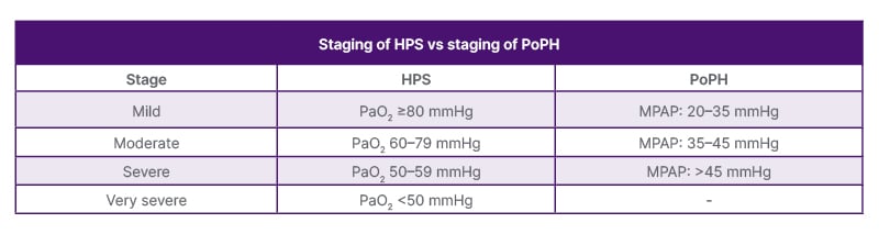
Table 1: Staging of HPS and PoPH.3-5
HPS: hepato-pulmonary syndrome; MPAP: mean pulmonary arterial pressure; PaO2: arterial partial pressure of oxygen; PoPH: porto-pulmonary hypertension; vs: versus.
CASE PRESENTATION
A female in her 60s with past medical history significant for primary biliary cholangitis (diagnosed via liver biopsy), autoimmune cirrhosis, mixed connective tissue disease, Sjögren’s syndrome, rheumatoid arthritis, and Raynaud’s phenomenon initially presented to the authors’ hospital with haematemesis and upper gastrointestinal bleeding. Family history was significant for liver cirrhosis in the mother and maternal grandmother. Surgical history was significant for appendectomy and cholecystectomy. Social history was significant for former tobacco use and occupation as a home health aide. The patient underwent variceal banding and a transjugular intrahepatic portosystemic shunt procedure during the first hospitalisation and did not require further hospitalisation until a COVID-19 infection several years later. During outpatient pulmonary follow-up in Month 1 of the second hospitalisation, peripheral capillary O2 saturation (SpO2) on room air was noted to be 94% during ambulation. Chest CT in Month 2 was negative for interstitial lung disease, with pulmonary function test (PFT) showing forced expiratory volume in 1 second/forced vital capacity 83.9%, forced expiratory volume in 1 second 100%, forced vital capacity 100.2%, total lung capacity 103.7%, and diffusing capacity of the lungs for carbon monoxide (DLCO) 63.2% of predicted values. In Month 9, the patient continued to endorse exertional dyspnoea of 6 months’ duration noticed when doing chores for more than 5–10 minutes and while walking short distances, and was therefore classified New York Heart Association Functional Classification (NYHA) III–IV by a referred cardiologist. Nuclear stress test during this time frame was negative for ischaemia. Transthoracic echocardiogram (TTE) showed left ventricular ejection fraction (LVEF) 65–70%, mildly enlarged left atrium, mild mitral regurgitation, mild tricuspid regurgitation, and pulmonary systolic pressure (PASP) of 45.5 mmHg. Right heart catheterisation was notable for systolic pulmonary arterial pressure (PAP) of 74 mmHg, mean pulmonary artery pressure (MPAP) of 41 mmHg, pulmonary capillary wedge pressure of 10 mmHg, and peripheral vascular resistance of 527.29 dynes-sec/cm5. The patient was then started on oral sildenafil and treprostinil for what was designated as Group I pulmonary arterial hypertension (PAH) during this period.
Following treatment initiation with oral prostacyclin and phosphodiesterase Type 5 (PDE5) inhibitors, repeat TTE in Month 16 showed mild pulmonary hypertension PASP of 47.9 mmHg with normal right ventricular (RV) size and function. A physical exam at this time was notable for digital clubbing and bilateral lower extremity oedema that was refractory to furosemide and spironolactone diuresis. The following TTE in Month 23 showed PASP of 43 mmHg with RV ejection fraction of 61%. The patient continued to follow up with gastroenterology, pulmonology, and cardiology, and subsequently presented to the hospital in Month 29 with a left frontal subarachnoid haemorrhage secondary to rupture of a developmental venous anomaly. Interval TTE conducted in Month 35 showed LVEF of 75% and PASP of 33 mmHg. During an outpatient visit with pulmonology in Month 38, SpO2 was 93% on room air at rest and 87% during a 6-minute walk test. Because the patient showed improvement (rest SpO2 of 98% and exertional SpO2 of 94%) while on a 2L nasal cannula, they were started on continuous O2 therapy.
The patient then presented to the authors’ hospital in Month 40 with shortness of breath and generalised weakness, and was discharged on higher doses of diuresis, only to return in Month 42 with similar concerns and SpO2 of 93% while on 3–4L home O2. Arterial blood gas during this hospitalisation demonstrated pH 7.5, partial pressure of CO2 in arterial blood (PaCO2) 39, PaO2 79, and bicarbonate 30 while on 2L O2 via nasal cannula, with chest radiograph showing severe scoliosis. Follow-up ECG with outpatient cardiology in Month 44 was notable for frequent premature atrial complexes and low voltage QRS complexes indicative of pulmonary disease, with patient SpO2 95% on 3L O2. In Month 45, contrast-enhanced microbubble TTE was ultimately positive for pulmonary venous shunt, with bubbles visualised in the left heart after six cardiac beats (Figure 1), LVEF of 69%, and PASP of 41 mmHg. CT angiography of the chest around the same time was notable for peripheral arteriolar dilatation with increased number of terminal branches extending towards the pleura, suggestive of HPS.
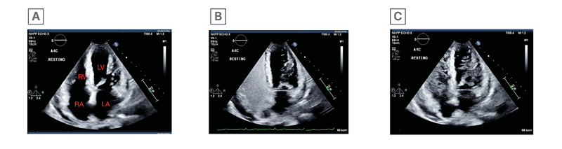
Figure 1: Trans-thoracic ecocardiogram apical four chamber view bubble study for case patient.
Trans-thoracic echocardiogram apical four chamber view bubble study demonstrating: (A) absence of bubbles before saline solution; (B) five cardiac cycles following saline microbubble introduction, with microbubbles appearing in the LV, indicative of an intrapulmonary shunt; and (C) 10 cardiac cycles following the injection of saline microbubbles, with microbubbles apparent in the LV. LA: left atrium; LV: left ventricle; RA: right atrium; RV: right ventricle.
Unfortunately, following one failed attempt at transplantation during which this patient became prohibitively hypoxaemic in the induction phase, there have not been further attempts of surgical intervention.
DISCUSSION
Interplay Between HPS and PoPH Pathophysiology
HPS is stratified into two types based on response to supplemental O2: Type 1, primarily caused by a ventilation perfusion mismatch resulting from increased O2 transit time to the central flowing red blood cells following dilatation of pre-capillary and capillary vessels to 15–100 μm diameters; and Type 2, primarily characterised by arteriovenous malformations (AVM) following cytokine-induced angiogenesis.8,9 In O2 supplementation responsive Type 1 HPS, the inadequate gas exchange at the red blood cell level coupled with an increase in the pulmonary capillary wall thickness in direct response to over-perfusion of the capillary bed leads to dampened values of DLCO, as seen in this patient case, with mildly reduced DLCO of 63.2% on PFT in Year 1.10 The mechanism of pulmonary vasodilatation characterising both types of HPS involves the activation of endothelial nitric oxide synthase by the annealing of endothelin-1 to pulmonary receptor enthothelin-1B in addition to the activation of inducible nitric oxide synthase via TNF-α inflammatory cytokines.6 The portal hypertension characteristic of cirrhosis allows for intestinal bacterial translocation to pulmonary vessels where macrophage activation of nitric oxide not only leads to further vasodilation through the haem oxygenase-carbon monoxide pathway, but also activates vascular endothelial growth factor angiogenesis implicated in the AVMs defining HPS Type 2.11 This patient’s history of cerebral venous anomaly, coupled with progression of autoimmune cirrhosis, predisposed them to the AVM intrapulmonary shunting detected via bubble study and categorises them with Type 2 HPS.
Predilection of AVM intrapulmonary shunt and increased perfusion with inadequate ventilation at the lung bases presents as progressive positional dyspnoea, or platypnoea, in the patient with HPS. Orthodeoxia or a decline in PaO2 of at least 4 mmHg via arterial blood gas or a measured 4% decline in SpO2 via pulse oximetry from supine to upright positioning is pathognomonic for HPS and for its umbrella term, platypnoea orthodeoxia syndrome.12 The clinical ramifications of this positional hypoxaemia consist of cyanosis, fatigue, spider nevi, and digital clubbing, with digital clubbing, a pertinent positive for this patient case, having a positive predictive value of 75% in HPS diagnosis.13
PoPH, on the other hand, develops in 2.0–8.5% of patients with portal hypertension, and is the result of increased pulmonary vascular constriction and resistance.14 Although ECG with agitated saline is gold standard for HPS vasodilatation and intra-pulmonary shunting detection, PoPH diagnosis hinges upon right heart catheterisation haemodynamics and is defined by: a) an MPAP >20 mmHg; b) a pulmonary capillary wedge pressure ≤15 mmHg; and c) pulmonary vascular resistance (PVR) >160 dynes-sec/cm5.5,15 As PoPH is primarily classified by MPAP (Table 1), this patient’s MPAP of 41 mmHg during initial exertional dyspnoea work-up placed them in the moderate PoPH category. Secondary to hyperdynamic vascular remodelling with arterial muscularisation and media hypertrophy, PoPH accounts for a considerable proportion of Group I PAH diagnoses, and, if inadequately addressed, can lead to increased RV afterloads and right heart failure.16 PAH therapies such as prostanoids, endothelin receptor antagonists, and PDE5 inhibitors are the designated treatment options for PoPH and are shown to improve pulmonary haemodynamics and 6-minute walk exercise capacity in these patients.17 The combination of PDE5 inhibitor and prostacyclin managed to reduce PASP to 24–33 mmHg from an initial 74 mmHg during interval haemodynamic surveillance of this patient via TTE.
While therapeutics such as pentoxifylline, methylene blue, and sorafenib targeting the pathophysiological factors underlying HPS have demonstrated efficacy in pilot studies, liver transplantation is the definitive treatment for HPS, and results in correction of hypoxaemia within 6–12 months.18 Demonstration of the HPS triad with PaO2 <60 mmHg bares prioritisation for transplantation, and patients meeting these criteria with a Model for End-Stage Liver Disease score of 22 are prioritised for this avenue of management.15 Elevated PAPs exceeding 50 mmHg in severe PoPH, however, contraindicate liver transplantation prior to PAH therapy in patients with PoPH. Liver transplantation following therapeutic directed decline in MPAP to <35 mmHg (mild staging of PoPH) has a reported 5-year survival rate of 67% and a 3-year survival rate of 77% in this population.19 Despite continued titration of the patient’s PoPH treatment with a regimen composed of sildenafil and intravenous epoprostenol following a repeat right heart catheterisation in Month 47, which revealed PASP elevated from 53 mmHg to 75 mmHg and mean PAP of 25–30 mmHg minimally vaso-responsive to intra-procedural nitric oxide, orthotopic liver transplantation (OLT) has been complicated by peri-operative hypoxaemia. Although the United Network for Organ Sharing (UNOS) allocates high priority to liver transplant candidates with HPS, this patient ultimately opted for the non-surgical approach to treatment and has been designated UNOS status 7 out of 40, with higher scoring indicating higher priority.20
Differential Diagnoses for PoPH and HPS Clinical Presentation
With the potential to present as a pulmonary complication of liver disease, hereditary haemorrhagic telangiectasia (HHT), also known as Osler-Weber-Rendu syndrome, is an autosomal dominant genetic condition that can be considered as a differential for both PoPH and HPS. In rare cases, HHT can present with diffuse AVMs that affect multisystem organs including the liver and lungs. Portal hypertension in HHT results from arteriovenous shunting between the hepatic artery and portal vein. Though primary symptoms of this condition include recurrent epistaxis and mucocutaneous telangiectasias, this diagnosis can present with acute hypoxaemia uncorrected with supplemental O2 administration and responsive to liver transplantation.21
Group V pulmonary hypertension related to autoimmune disease/mixed connective tissue disease is an additional diagnosis on the differential, as its pathophysiology mimics PAH caused by progressive vascular remodelling and constriction that is present in <1% of mixed connective tissue disease. PFTs reveal restrictive patterning and reduced DLCO with this pathology, as demonstrated by the case patient.22
The hyperdynamic state in the setting of cirrhosis is characterised by an increased circulatory volume coupled with a decreased systemic vascular resistance with a cardiac output (CO) greater than 8 L/min calculated via direct Fick methodology. Increased pulmonary circuit blood flow and shear stress compounded by increased left ventricular end-diastolic pressure causes pulmonary vascular remodelling through the release of vasoactive molecules that lead to vasoconstriction. In cirrhosis, the hyperdynamic state results from splanchnic vasodilation, decreased effective blood volumes, and reflexive activation of the renin-angiotensin-aldosterone system. PVR is low to normal and wedge pressure is normal to high, with liver transplantation again being the definitive treatment for this hyperdynamic state.23
Pre-operative Management of Patients with PoPH and HPS
Thirty-day mortality of patients with pulmonary hypertension undergoing elective non-cardiac and non-obstetric general surgeries lies between 2–18%, and most often results from acute RV failure. In patients with PAH or PoPH undergoing liver transplantation, mortality increases to 35%.24 RV dysfunction characterised by chamber enlargement, reduced tricuspid annular plane systolic excursion, and reduced ventricular fractional shortening portend greater pre-operative risk in the patient with pulmonary hypertension. MPAP >45–40 mmHg and PVR >400 dynes-sec/cm⁵ are also absolute contraindications to liver transplantation.5 It is therefore crucial to discuss pre-operative planning and risks, including an a priori anaesthetic management plan in a multidisciplinary team consisting of pulmonary hypertension specialists, anaesthesiologists, transplant surgeons, intensivists, and extracorporeal membrane oxygenation (ECMO) operators.
Pre-operative management entails an individualised pre-operative risk assessment, as well as medication optimisation with endothelin receptor antagonists, PDE5 inhibitors (such as sildenafil 20–40 mg ter in die dosing), calcium channel blockers, and prostanoids prior to surgery. Elevated peri-operative risk is determined by patient factors such as RV dysfunction, as measured by right atrial pressure >7 mmHg, PVR >128 dynes-sec/cm⁵, and MPAP >35 mmHg; exercise intolerance measured by 6-minute walk distance <400 m; and WHO functional status III/IV, in addition to procedural factors such as duration >2–3 hours.5,24 Cardiopulmonary exercise testing evaluating peak O2 uptake and end tidal CO2 tension offers additional insight into risk prognosis in patients with PoPH. Titration of pulmonary vasodilators to WHO functional class, 6-minute walk distance, right arterial pressure, MPAP, and PVR is necessary prior to incision, with therapies continued uninterrupted peri and intra-procedurally. Epoprostenol has a half-life of 3–5 minutes and must be administered intravenously via an indwelling catheter in the pre-operative setting. Treprostinil is a more stable prostacyclin compound and can be administered subcutaneously, while iloprost is a stable prostacyclin analogue with a half-life of 20–30 minutes that is administered via inhalation at lower doses.25 Intermittent nebulised prostacyclin, continuously inhaled epoprostenol, inhaled nitric oxide, or intravenous prostanoids should be considered for continuous administration both intra-operatively and post-operatively.
Anaesthesia Considerations for Liver Transplantation in Patients with PoPH
Cistacurium has liver independent elimination, as opposed to vecuronium and rocuronium, and should therefore be the paralytic agent of choice during OLT.25 Anaesthetic drugs directly impact cardiac muscle calcium cycling on the cellular level, leading to autonomic reduction in contractility.24 Inhaled anaesthetics such as isoflurane and desflurane decease adenosine triphosphate-dependent potassium channels needed for vasodilation and therefore increase RV afterload. These volatile anaesthetic agents can be administered in up to one minimum alveolar concentration without disturbing pulmonary vasculature resistance.26 Though correlated with adrenal suppression, etomidate has haemodynamic stability and is used in some operations with patients who have pulmonary hypertension as it has minimal effect on systemic vascular resistance (SVR), PVR, and contractility.24 Nitrous oxide and protamine may increase PVR and should be avoided in this subset of patients. Although ketamine decreases PVR minimally, it has sympathetic effects that increase RV contractility and SVR to counteract the vagal responses of general anaesthesia that are beneficial in OLT. During induction, propofol can be used along with a vasopressor to avert hypotension. Vasopressors norepinephrine, vasopressin, and phenylephrine maintain coronary perfusion in the setting of hypotension caused by the various induction agents. In very high-risk cases, ionotropes dobutamine and dopamine, coupled with pressors, can be started prior to induction with continuation postoperatively. ECMO vascular sheaths can additionally be inserted during induction to allow for efficient transfer to this modality if a need for RV offloading post-procedurally is anticipated.
After the airway is established with targeted pre-oxygenation, ventilator management of the patient with PoPH is geared towards higher fraction of inspired O2, hyperventilation to sustain PaCO2 of 30-35 mmHg without pH <7.4, positive end expiratory pressure (PEEP) between 5–10 cmH20, and lung tidal volumes at normal functional residual capacity. High plateau pressures, tidal volumes, and PEEP >5 mmHg during mechanical ventilation compresses intra-alveolar vasculature and should be averted as to limit PVR and RV dysfunction.27 Intermittent positive pressure ventilation in conglomerate with endogenous PEEP and abdominal insufflation increases RV afterload and decreases right coronary perfusion.26
Since PAP is equivalent to the product of CO and PVR summed with left atrial pressures (PAP=[COxPVR]+left atrial pressures), RV failure is evidenced by both PVR and RV filling pressure increases with concomitant CO decreases.28 When large increases in PVR and RV afterloads are experienced, right ventriculo-arterial uncoupling occurs intra-operatively and leads to systemic hypotension. Coronary blood flow to the RV is decreased during systole if pressure within the cavity is higher than or equal to systemic pressures. RV failure furthermore occurs when wall stress and endomyocardial pressure approaches aortic pressure and leads to threatened systolic RV coronary blood flow. RV function in PoPH is fluid dependent. Maintenance of adequate RV preload with fluids or blood products requires precise titration, as an excessive increase can lead to tricuspid regurgitation and ventricular dysfunction, while excessive decrease due to blood loss, diuresis, intra-abdominal pressure, and vena cava compression can cause low CO and hypotension.24 Intra-operative hypotension and decreased CO should be managed with vasopressor therapy rather than downtitration of intravenous or inhaled pulmonary vasodilators. Milrinone inhibits cyclic adenosine monophosphate breakdown, enhances myocardial contraction, reduces systemic and pulmonary vascular tone, and counteracts the right ventricle’s inability to handle the acute increase in CO seen in the reperfusion stage of OLT. Given that iono-vasodilators such as dobutamine (2–5 µg/kg/min) and milrinone (50 µg/kg bolus and 0.5–0.75 µg/kg/min) allow for CO maintenance at the expense of reduced SVR, inhaled nitric oxide at dose 5–20 ppm, inhaled iloprost at 5–10 µg, or intravenous prostacyclin/sildenafil should be employed in response to RV failure-induced systemic hypotension.29,30 Vasopressors should be used in conjunction to prevent RV ischaemia, and preload should be optimised with fluid challenges or diuresis during pulmonary hypertension crises. Vasopressin in particular causes systemic vasoconstriction coupled with pulmonary vasodilation via nitric oxide release, and is the pressor of choice during the pre-anhepatic and anhepatic phases of OLT.31 In the event of overt RV dysfunction, which can occur during the pre-anhepatic phase due to an acute blood loss, during the anhepatic phase due to clamping of the inferior vena cava, or during the reperfusion phase due to cytokine storm, ionotropes norepinephrine and epinephrine should be utilised.31 Intraoperative haemodialysis, diuresis, nitroglycerin bolusing, veno-arterial VA ECMO, or percutaneous RV assist devices may also be used to offload the RV in high-risk cases.24
Intra-operative management entails continuous pulmonary artery pressure monitoring via pulmonary artery catheter, stroke volume monitoring via arterial pulse contour analysis, and RV function monitoring with transoesophageal echocardiogram (TEE) or with central venous pressure monitoring if skilled TEE is unavailable.29 Studies demonstrate a 30–50% significant change in therapeutic management in patients being monitored with TEE during non-cardiac surgery. The aim of anaesthesia management is to maintain right ventricular CO and to minimise systemic hypotension with vasopressor support. As hypotension can cause rapid RV failure via ischaemia, continuous blood pressure monitoring with an arterial catheter is also paramount.
In the postoperative arena, invasive monitoring should be continued in the ICU for at least 48–72 hours with uninterruption of PoPH therapies. PDE5 inhibitors allow for weaning from inhaled pulmonary vasodilators.29 In this stage of treatment, hypothermia causes pulmonary vasoconstriction and ventilation perfusion mismatch so analgesia is needed to temper sympathetic overdrive and to reduce increases in PVR. Additional conditions that contribute to increased PVR, such as hypoxaemia, hypercapnia, acidosis, and hypervolaemia, should be curtailed in the postoperative setting.30 Early extubation is optimal in order to limit intrathoracic pressure and RV stress. Hypoxaemia may persist for several weeks to a year following transplantation in a patient with HPS, in which case non-invasive positive pressure ventilation can be used as a bridging therapy. Postoperative deaths in the case of patients with PoPH who have undergone liver transplants are attributed to fluid shifts, pulmonary vasoconstriction, arrhythmias, and pulmonary thromboembolism.25
CONCLUSION
In conclusion, the authors report a case of cirrhosis that developed PoPH. During the treatment for PoPH with pulmonary vasodilator therapy, the patient developed worsening hypoxaemia requiring supplemental O2. A TTE with agitated saline bubble study showed extensive right-to-left shunt with visible arteriolar dilatations on contrast-enhanced CT chest, suggesting diagnosis of HPS similar to a case reported in Germany.32 Physicians should be aware of this transition of two competing mechanisms in the same patient that completely changes treatment strategies in such patients. Liver transplantation, the definitive treatment for HPS, requires multidisciplinary planning in addition to close intra-operative monitoring at the behest of skilled anaesthesiologists. Pre-operative management entails an individualised pre-operative risk assessment, as well as medication optimisation with endothelin receptor antagonists, PDE5 inhibitors, calcium channel blockers, and prostanoids. Anaesthesia considerations during OLT overall aim to minimise PVR, avert RV ischaemia by maintaining preload-dependent coronary perfusion pressures, and prevent RV failure by maintaining coupling between the RV chamber and the pulmonary arterial vasculature.