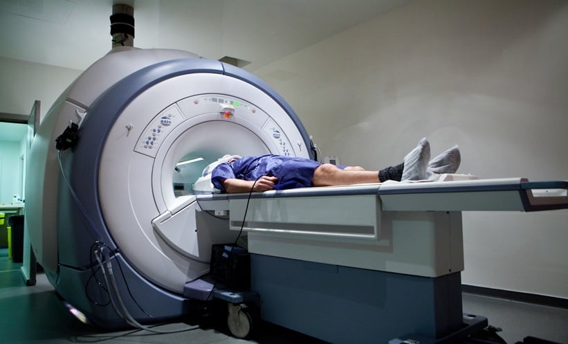
DISTINGUISHING healthy tissues from cancerous tissues in prostate cancer patients with a high degree of specificity and sensitivity has been shown to be possible by the results of a recent study. This is an important step in the management of prostate cancer, as it will enable early diagnosis and thus increase the efficacy of treatment. However, despite recent advances diagnosing methods, it can still be challenging to successfully diagnose prostate cancer. The study’s lead author, Dr Vikas Prasad, Charité–Universtätsmedizin Berlin, Berlin, Germany, commented on this difficulty, stating: “However, in many patients, histopathology may not yield correct diagnosis (e.g., if the tumour is missed during true-cut biopsy). This is especially true for multifocal prostate cancer, less aggressive tumours, and cases of prostatitis or prior prostate irradiation, where magnetic resonance imaging (MRI) alone may not give the correct localisation and malignancy grade.” Therefore, this work, which is believed to be the first to determine a cut-off maximum standardised uptake value (SUVmax) validated by immunohistochemistry, is exceedingly valuable.
In this retrospective study, the authors set out to determine a cut-off SUVmax that would identify prostate cancer. The study cohort comprised 31 males with a mean age of 67.2 years who had previously undergone preoperative positron emission tomography (PET) scans and prostatectomies. A combined total of 62 cancerous and benign prostate tissue samples were stained with monoclonal antiprostate-specific membrane antigen. It was possible to confirm all cancerous tissues histopathologically. An analysis was then conducted to determine the optimum SUVmax cut-off value; this was found to be 3.15 (sensitivity: 97%; specificity: 90%). This cut-off value will facilitate the diagnosis of prostate cancer in both unifocal and multifocal disease with a high degree of specificity and sensitivity.
Speculating about the future, Dr Prasad suggested: “With the advancement of image-registration and segmentation software and PET/magnetic resonance imaging (MRI) scanners, it is quite logical to predict that in the future PET images and SUVmax on a suspicious lesion in the prostate will be used for multimodal image-guided fusion biopsy.”