BACKGROUND
Ischaemic injury of the adult human heart results in irreversible loss of cardiomyocytes (CM), leading to fibrosis, and eventually heart failure. Cardiac regeneration attempts involving the transplantation of somatic progenitor cells uniformly failed to provide relevant de novo CM and did not reverse heart failure in multiple clinical trials.1,2 As an alternative method of CM provision, direct reprogramming of fibroblasts into induced CM (iCM) by forced expression of cardiomyogenesis-related transcription factors is currently being explored. However, reprogramming efficiency is low, and mature iCM have little or no proliferation capacity. The authors therefore developed a protocol for the genetic reprogramming of cardiac fibroblasts (CF) into iCM precursor cells (iCMP) with CM lineage commitment and evaluated their therapeutic potential in a mouse model of myocardial infarction (MI).
METHODS AND RESULTS
CF were reprogrammed into iCMP by lentiviral gene transfer of GATA4, MEF2C, TBX5, and MYOCD (GMTMy),3 resulting in approximately 30% of cells positive for cardiac troponin T, α-actinin, and myosin heavy chain in immunostainings. The induced cells also expressed well-known cardiovascular precursor markers such as Nkx2-5, Mesp1, CXCR4, and Flk-1, and displayed robust proliferation capacity as indicated by Ki67 expression. Next, pure iCMP populations were obtained by transcriptional selection with MYH6/7-targeting molecular beacons.4
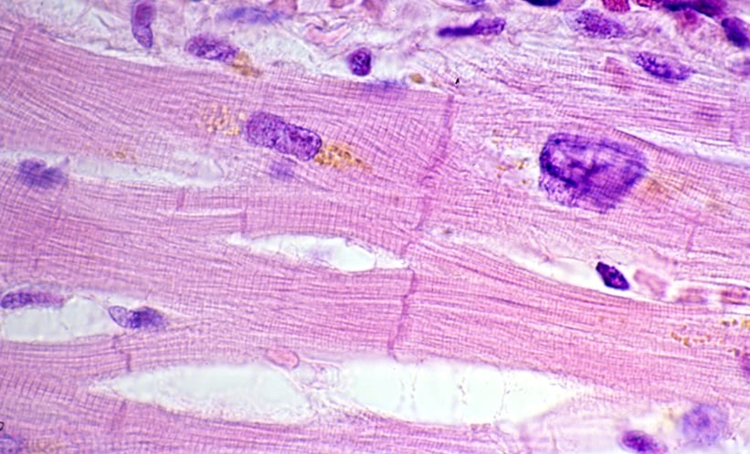
Thereafter, global transcriptome profiling was performed by RNA sequencing. By principal component analysis, iCMP displayed a unique gene expression profile compared to their parental CF and adult heart, indicating an intermediate state of cardiac development. Gene Ontology analyses revealed upregulation of genes associated with cardiac development, differentiation, and morphogenesis, as well as downregulation of genes associated to cell proliferation in iCMP compared to CF. Evaluation of selected gene sets showed downregulation of nonmyocyte genes, upregulation of additional cardiac transcription factors, and upregulation of certain functional and structural genes, such as ion channel genes and contractile genes. Subsequently, iCMP were differentiated towards CM using 5-Azacytidine, TGFß, and ascorbic acid. In these differentiation assays, iCMP ceased proliferation, displayed elongated morphology, and formed prominent sarcomere-like structures. CD31-positive endothelial cells were not detected in differentiating iCMP cultures, but the presence of α-smooth muscle actin-positive smooth muscle cells was noted. iCMP-derived CM did not display spontaneous contractions within the 20-day-observation period.
To generate sufficient cell doses for intracardiac injection in a mouse model of MI, iCMP were extensively expanded in medium supporting their stable phenotype. Frozen stocks were prepared and iCMP phenotype was confirmed after thawing and recovery before in vivo application in male C57BL/6J mice after permanent ligation of the left anterior descending artery. Serial transthoracic echocardiographic analyses revealed that left ventricular ejection fraction was increased as early as 2 weeks after iCMP injection, compared to the no treatment and placebo control groups. This increase was stably maintained until Week 6. Similarly, cardiac output was improved 6 weeks after iCMP treatment. Additionally, an improvement in left ventricular wall thickness at diastole in mice transplanted with iCMP was observed, as well as a dramatically decreased scar size in histological sections stained with Masson’s trichrome.
CONCLUSION
Taken together, iCMP generated via direct cellular reprogramming followed by transcriptional selection display a phenotype compatible with an intermediate state of cardiogenic development. They can be expanded to yield therapeutic cell doses and beneficially influence post-infarct myocardial remodelling in a rodent model. Thus, iCMP are promising candidates for novel cardiac cell therapy/regeneration strategies.