Abstract
Background: Cholesterol pericarditis is a rare form of pericardial disease characterised by chronic cholesterol-rich pericardial effusions, commonly associated with rheumatoid arthritis (RA), hypothyroidism, or tuberculosis. These effusions may progress to cardiac tamponade, requiring urgent intervention.
Case Presentation: A 66-year-old male with RA presented with fatigue and dyspnoea and was found to have a large pericardial effusion with echocardiographic evidence of cardiac tamponade. Initial pericardiocentesis drained 600 mL of cholesterol crystal-containing fluid; however, rapid reaccumulation required repeat drainage with temporary catheter placement. Pericardial fluid analysis demonstrated elevated cholesterol (80 mg/dL), markedly elevated lactate dehydrogenase (>2500 U/L), and inflammatory cell predominance, with negative cultures and cytology. The patient was managed with pericardiocentesis, drainage, colchicine, and continuation of RA-directed therapy without surgical intervention. Over 1 year, serial echocardiography demonstrated normalisation of cardiac function and complete resolution of the effusion.
Conclusion: This case highlights that conservative, non-surgical management may be effective in selected patients with RA-associated cholesterol pericarditis complicated by tamponade. A structured diagnostic approach and individualised management strategy are critical in optimising outcomes.
Key Points
1. Cholesterol pericarditis is a rare inflammatory pericardial condition often linked to rheumatoid arthritis, characterised by chronic, cholesterol-rich pericardial effusions that can cause cardiac tamponade requiring urgent intervention.2. While pericardiectomy is typically recommended for definitive management of cholesterol pericarditis, this case demonstrates that careful conservative management with pericardiocentesis, drainage, and medical therapy can successfully resolve tamponade and prevent recurrence.
3. Close multidisciplinary follow-up with cardiology and rheumatology is critical in managing cholesterol pericarditis, allowing for tailored treatment approaches and monitoring of cardiac function and effusion recurrence over time.
INTRODUCTION
Pericarditis is the most common form of pericardial disease encountered in clinical practice and may occur with or without pericardial effusion.1,2Cholesterol pericarditis is a rare subtype characterised by chronic, cholesterol-rich pericardial effusions and the presence of cholesterol crystals within the pericardial fluid. It has been associated with chronic inflammatory conditions such as rheumatoid arthritis (RA), as well as hypothyroidism and tuberculosis.3
The pathogenesis is thought to involve chronic inflammation leading to cellular membrane breakdown and accumulation of cholesterol within the pericardial space. These effusions are often long-standing and may exhibit the classic ‘gold paint’ appearance. Although typically indolent, cholesterol pericarditis can progress to cardiac tamponade or constrictive pericarditis.
The authors present a case of RA-associated cholesterol pericarditis presenting with cardiac tamponade that was successfully managed with a non-surgical approach, along with a focused review of current management strategies.
CASE PRESENTATION
A 66-year-old male with a 2-year history of RA presented to the emergency department after an outpatient transthoracic echocardiogram (TTE) revealed a large pericardial effusion (Figure 1). He reported several days of progressive fatigue and dyspnoea. Three days prior, a chest radiograph obtained for fatigue demonstrated cardiomegaly, prompting urgent echocardiographic evaluation.
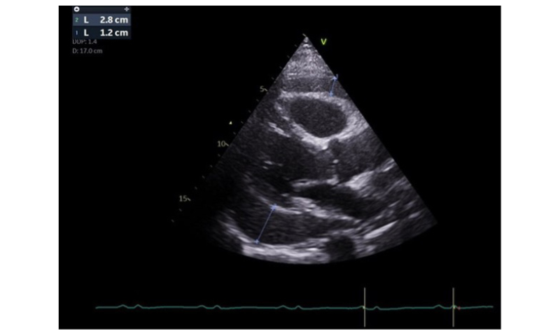
Figure 1: Transthoracic echocardiogram demonstrating a large circumferential pericardial effusion appearing as an anechoic (dark) space surrounding the heart.
On presentation, vital signs were notable for blood pressure 158/93 mmHg, heart rate 83 beats per minute, respiratory rate 15 breaths per minute, oxygen saturation 98% on room air, and temperature 98.6 °F. Physical examination revealed muffled heart sounds without jugular venous distension or peripheral oedema.
ECG demonstrated normal sinus rhythm with low voltage in the extremity leads.
TTE revealed a large circumferential pericardial effusion measuring up to 2.8 cm with reduced left ventricular ejection fraction (LVEF) of 45–50%. Echocardiographic findings consistent with cardiac tamponade included right atrial and right ventricular diastolic collapse and a plethoric inferior vena cava.
Laboratory studies demonstrated total cholesterol of 165 mg/dL, high-sensitivity troponin of 28.6 ng/L, erythrocyte sedimentation rate of 31 mm/hr, and C-reactive protein of 3.2 mg/dL. Thyroid-stimulating hormone was normal at 2.1 µIU/mL.
The patient’s RA had been managed with methotrexate 15 mg weekly, prednisone 20 mg daily, and etanercept 50 mg weekly, all of which were continued during hospitalisation.
Urgent pericardiocentesis was performed under sterile conditions with fluoroscopic and echocardiographic guidance. A total of 600 mL of cloudy yellow fluid was removed. A pericardial drain was briefly placed but removed after confirmation of near-complete drainage, as reaccumulation was not initially anticipated.
Pericardial fluid analysis demonstrated:
- white blood cell count: 1,888/µL;
- red blood cell count: 900/µL;
- lactate dehydrogenase: >2500 U/L;
- albumin: 2.2 g/dL;
- cholesterol: 80 mg/dL; and
- microscopy: numerous cholesterol crystals.
Gram stain, bacterial cultures, and blood cultures were negative. Cytology demonstrated no malignant cells. Triglycerides and glucose were not obtained.
Despite initial improvement, serial echocardiography demonstrated rapid reaccumulation of the effusion (Figure 2). A second pericardiocentesis was performed, removing an additional 200 mL of fluid, and a pericardial drain was left in place. Drain output decreased over 2 days (6 mL, then 10 mL), and follow-up imaging showed only trivial residual effusion, after which the drain was removed.
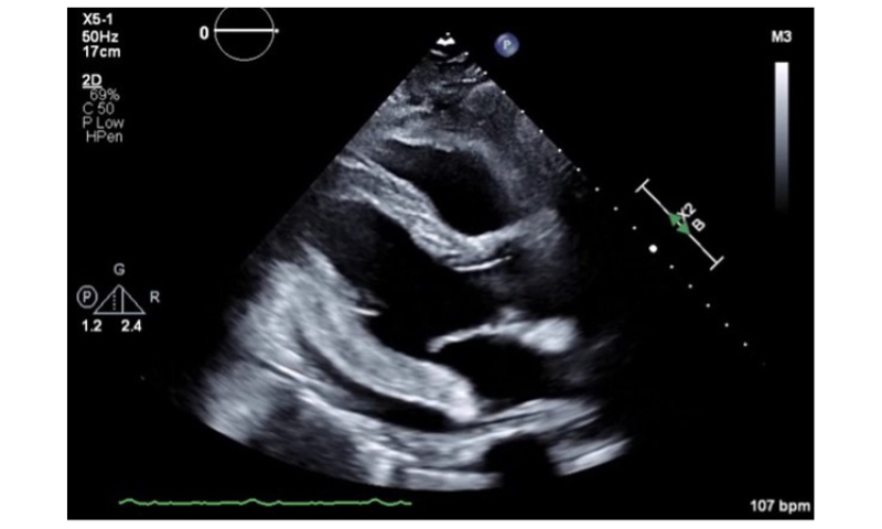
Figure 2: Transthoracic echocardiogram following initial pericardiocentesis, showing a residual small to moderate pericardial effusion.
The patient was discharged on colchicine (1.2 mg twice daily on Day 1, followed by 0.6 mg twice daily, planned for 3 months). Nonsteroidal anti-inflammatory drugs were not used, and corticosteroids were continued as part of RA management without adjustment.
At 2-week follow-up, TTE demonstrated preserved LVEF (55–60%) and trivial effusion. Serial imaging over 1 year showed progressive improvement, with LVEF increasing to 65–70% and complete resolution of the pericardial effusion (Figure 3). No recurrence or complications were documented.
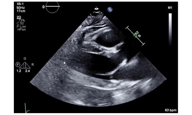
Figure 3: Transthoracic echocardiogram at 1-year follow-up showing complete resolution of pericardial effusion.
DISCUSSION
Diagnostic Considerations
Cholesterol pericarditis is defined by the presence of cholesterol crystals within pericardial fluid, often accompanied by elevated pericardial fluid cholesterol levels (commonly >70 mg/dL) and chronic effusion.4 In RA, the high cholesterol concentration is thought to be from release of cholesterol from injured pericardial cell membranes or necrotic rheumatoid nodules.5 It has been observed that the pericardial fluid often has a characteristic ‘gold paint’ consistency.6
In this case, the diagnosis was supported by the presence of cholesterol crystals, elevated pericardial fluid cholesterol, and an inflammatory effusion. Alternative aetiologies were systematically excluded. Infectious causes were considered unlikely given negative blood and pericardial fluid cultures. Tuberculosis was deemed low probability due to lack of risk factors or clinical features. Hypothyroidism was excluded with normal thyroid function testing, and cytology was negative for malignancy.
The association with RA, along with chronic inflammation and absence of alternative causes, supported RA-associated cholesterol pericarditis.
Aetiology and Pathophysiology
Cholesterol pericarditis is most commonly associated with chronic inflammatory conditions such as RA, though it may also occur in tuberculosis, hypothyroidism, or idiopathically.3,7,8 Chronic inflammation is thought to promote breakdown of cellular membranes, leading to cholesterol accumulation and crystal formation within the pericardial space.
Management Strategies
Initial management involves pericardiocentesis, which is both diagnostic and therapeutic, particularly in cases of cardiac tamponade. However, recurrence is common, and definitive management has traditionally involved pericardiectomy.3,9 Without it, there is a risk of recurrent effusions, which can lead to progressive pericardial thickening and scarring, ultimately increasing the risk for constrictive pericarditis over time.10
Surgical intervention is generally recommended for:
- recurrent or persistent effusions;
- development of constrictive physiology; or
- failure of conservative management.
Less invasive options such as pericardial window may be considered in selected cases.
Role of Medical Therapy
The role of medical therapy in cholesterol pericarditis is not well defined. Colchicine is commonly used in acute and recurrent pericarditis and may reduce inflammation and recurrence, though data specific to cholesterol pericarditis are limited. Nonsteroidal anti-inflammatory drugs and corticosteroids may also be used depending on the underlying aetiology, particularly in autoimmune disease.
In this case, colchicine was used in conjunction with continued RA-directed therapy, and no recurrence was observed over 1 year.
Monitoring and Risk of Constriction
Patients with cholesterol pericarditis are at risk for progression to constrictive pericarditis due to chronic inflammation and fibrosis.10 Close follow-up with serial echocardiography is essential to monitor for recurrence and early signs of constriction, such as ventricular interdependence or septal bounce.
Clinical Implications
This case demonstrates that even in the presence of cardiac tamponade and early recurrence, a conservative approach incorporating repeat drainage and medical therapy may be effective in selected patients. Careful patient selection and close multidisciplinary follow-up are critical.
Strengths and Limitations
A major strength of this report is the detailed clinical characterisation and 1-year follow-up demonstrating sustained resolution without surgical intervention. Limitations include incomplete pericardial fluid characterisation (e.g., triglycerides, glucose) and limited long-term follow-up beyond 1 year.
CONCLUSION
Cholesterol pericarditis is a rare but important cause of chronic pericardial effusion that may present with cardiac tamponade. This case illustrates that conservative management with pericardiocentesis, drainage, and adjunctive medical therapy can be effective in selected patients. Individualised management and close follow-up are essential to optimise outcomes and monitor for recurrence or progression to constrictive disease.
Patient Perspective
The patient reports significant improvement in symptoms and has experienced no recurrence. He tolerated therapy well and expressed satisfaction with the outcome of his care.