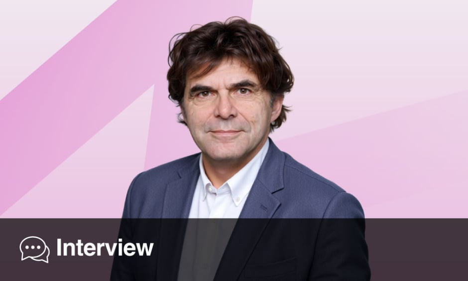
Harald Kittler | Professor, Department of Dermatology, Medical University of Vienna, Austria
Citation: EMJ Dermatol. 2026; https://doi.org/10.33590/emjdermatol/KEIZ706J.
How has dermatoscopy changed the way dermatologists approach the diagnosis of pigmented lesions?
It has changed the approach very much. Dermatoscopy became popular in the 1990s with the invention of the handheld dermatoscope. Before that, melanoma was often diagnosed later, when the disease was obvious to the unaided eye. Dermatoscopy shifted the diagnosis towards earlier disease. We now have an instrument that is small, easy to use, and cheap. It’s also efficient and easy to learn. It changed the landscape of melanoma diagnosis in the 1990s, firstly for dark, pigmented lesions, but then gradually expanded to non-pigmented lesions, including inflammatory dermatosis in the later years. The evolution has been towards a more general instrument for dermatology, like the stethoscope is for internal medicine.
What have been the most important developments in dermatoscopy over the past 2 decades, and how have they influenced clinical practice?
We have to first differentiate between instrumental and procedural improvements. I think the most important one was the invention of the handheld dermatoscope. As I already said, this changed the landscape, because if dermatoscopy required a large, cumbersome, expensive instrument, the success would not have been the same. Second, we learned to interpret what we saw under the dermatoscope over the years, and we came up with rules in a deductive system that allowed us to create algorithms to improve the diagnosis of all kinds of skin diseases. With more experience, we moved away from the algorithms to making a top-down, inductive, very quick diagnosis, because we got used to the patterns we saw with the dermatoscope. So, I would say the most important developments were the instrument, then the method, and because we used these for a long time, we became very good at recognition.
Now we have additional hardware, and also some procedural improvements. Hardware-wise, we have digital dermatoscopy that allows sequential imaging. We also have the inclusion of AI, and we have a more complex landscape of non-invasive imaging techniques where dermatoscopy is just a part of it, such as optical coherence tomography or confocal microscopy. We also have total body photography where dermatoscopy is integrated, now in conjunction with AI. I think the last big step was the use of dermatoscopy algorithms for inflammatory diseases and non-pigmented or neoplastic lesions.
The newest approaches and hardware improvements are AI and high-magnification dermatoscopy. We now also have additional lighting sources like UV and the popularisation of polarised dermatoscopy, which improved the way we look at pigmented and non-pigmented lesions.
What are the key principles clinicians should understand when learning dermatoscopy?
The rules are easy, and what you see is easy. We made it too hard in the past by making the morphology and terminology too complicated. It presented dermatoscopy as difficult to learn, which it is not. So, I would say in principle, it’s a bottom-up method. When you use pattern analysis, which is an all-inclusive and very simple method, it can be taught in 5 minutes. You have to use it for a long time to become an expert, but the basics can be learnt very quickly. I would say the most important advice is just to learn the basic rules, consider what you see and what you don’t see, and then keep using it and get better through experience. A beginner’s mistake is to use it five times a day, but you have to use it 100 times a day.
What are some of the most common pitfalls clinicians encounter when interpreting dermatoscopic images?
First of all, dermatoscopic images are not 100% accurate. You have to deal with the mistakes and limitations of the technique and integrate it with clinical information like anatomic side and age, as well as additional non-invasive examination. Dermatoscopy is a part of an ecosystem of aids to come to a diagnosis. Sometimes we cannot come to a diagnosis with certainty, have to remove the lesions, and then ask the pathologist. Sometimes not even the pathologist can come to a conclusion with certainty. This is the main obstacle that medicine has to face in general, but is particularly present in diagnostic techniques like dermatoscopy.
The second thing is that it takes a little bit of a learning curve. Of course, the basics are easily understood, but calibrating yourself to find your own way of doing dermatoscopy and getting experience is what you need to interpret the findings in a reasonable manner. This takes some time, and there is no easy way. You can’t bypass that. You cannot go to a course over a weekend and then know how to do dermatoscopy. There’s an inductive part that is based on your experience. This takes some time, so that’s one of the major obstacles.
How important is clinical experience in dermatoscopy, and what can structured diagnostic algorithms add to the decision-making process?
You always start to learn a method by learning the rules, like when we learn grammar. On the other hand, we learn to speak just by listening to others and repeating. Algorithms and deductive rules are an important top-down method to interpret these, like a ladder that lets you take steps to come to a specific diagnosis by applying rules. However, that has limitations because, first, you can still reach the wrong conclusion even when you apply the rules correctly. Secondly, it has to become implicit and intuitive. In the beginning, you use the rules explicitly, but to become faster, you need to make them so implicit that you just know them. When you become more experienced, you don’t need the ladder every time you have a problem. Once in a while, though, you will meet a lesion you can’t identify just by pattern recognition, so you need to take out the ladder again and use an algorithm or established method.
How should clinicians integrate dermatoscopic findings with a patient’s clinical history and individual risk factors?
There are many ways to integrate dermatoscopic findings. The weighting can change depending on the situation. Sometimes age or history are very important pieces of information, but sometimes they’re not. This makes it more complicated and complex. It also depends on where you practise dermatoscopy and whether you practise in a hotspot of melanoma, like Queensland, Australia, or in another part of the world where melanoma is very rare. There is no golden rule for how to weigh the context, or even how to weigh the different features you see with dermatoscopy.
You have to find your own way to do it. Deciding whether you should biopsy something or not depends on your personal threshold. You could have a low threshold, meaning you have high sensitivity and don’t overlook many melanomas or malignant lesions. Alternatively, you could have a high threshold, meaning you don’t have a lot of false positives. This also depends on your personal way of practising medicine. Depending on this, you will give different weight to different information. Some believe that history shouldn’t be weighed at all. This is a very personal decision, because some clinicians may see history as unreliable in the case of a 50-year-old being asked about a lesion on their back, but as reliable in the case of a 30-year-old asked about a lesion on their face. It depends on so many things, and it’s more of an art than a science.
Dermatoscopy is increasingly being used beyond pigmented lesions. Do you think it should become a routine component of dermatological examination more broadly?
Dermatoscopy is a routine component in many places already, but there are some parts of the world where it isn’t. In the past, we neglected individuals with dark skin because melanoma or skin cancer is more prevalent in individuals with fair skin, but this is now over. We know about our mistakes in the past, and there are books that deal only with dermatoscopy in dark skin, where neoplastic diseases are not so much a problem, but more inflammatory diseases like lupus, psoriasis, and lichen planus are.
Considering that dermatoscopy is useful for all skin types and in inflammatory diseases, there should be no part of the world where dermatoscopy is not part of the routine examination. Even in parts of the world where skin cancer is of low incidence, there is no reason not to use dermatoscopy.
There are still obstacles, though, because the dermatoscope is still too expensive in some parts of the world. Ideally, this would not be the case, and every doctor would have a dermatoscope, just as every cardiologist has a stethoscope. The fact that dermatoscopes are not routine is an inequality, not because dermatoscopy is not useful in some parts of the world.
What role do you see AI playing in dermatoscopic diagnosis, and how might it complement the expertise of dermatologists?
AI is a double-edged sword. There are the doomers and the bloomers. The doomers say we will not learn anything anymore and will be replaced by AI. The bloomers say it’s the solution to everything because we won’t need to learn dermatoscopy anymore. I’m in the middle because AI currently is just an instrument. There’s no agency. The agency comes from the person who holds the dermatoscope, or the hardware that is equipped with AI, so either way, it is our responsibility. We can benefit from the use of AI. It can make us smarter.
It is a complex relationship. You always have to decide how to use an instrument. There are things that are less important that we can use AI for, and then use our brains for better things. It is easy to use AI to compare lesions, to find the ones that have changed when a patient has hundreds of them. That is difficult for humans. Also, if you want to have a second opinion very quickly, AI is useful. On the other hand, it’s an instrument that is not 100% accurate. There is a tendency to assume it works all the time, but it doesn’t. We as humans constantly need to check if the tool we use is still accurate, and currently only we can check if it is. When you use a large language model and ask it something, you have to be critical towards the output. You cannot copy and paste. It is the same for the use of AI in dermatoscopy. It’s useful, but it also needs a smart person using it. I believe it can make smart people smarter.
What emerging developments in imaging technology are you most excited about?
In dermatoscopy, there are currently two emerging technologies. One is UV dermatoscopy, which can be useful in some very specific applications. For example, finding a mite, ruling out tinea, or diagnosing psoriasis. Some say it could also increase our accuracy for melanoma, but I’m a little bit critical there. Then there is high magnification dermatoscopy, which is cool because you can get a magnification near cellular level. It looks a little bit like confocal images. However, nobody knows if it’s really that useful. . Regarding AI, I think it is the third most promising technology that is evolving, and we’re still looking at ways to integrate it into practice. Although AI is useful, we don’t know how to best integrate it into our workflow. Should we always use it? Should we use it only if we are unsure? Should we have an AI-in-the-loop system like a co-pilot?
Looking ahead, what do you think the dermatoscopy of the future will look like?
I think it will not be much different from how we do things now. We will still have a handheld dermatoscope. The question is, who will be using it? Only dermatologists, or general practitioners and other medical personnel too? The dermatoscope itself will, of course, evolve, but it’s already rather optimal. It’s small and cheap, but maybe it could be equipped with AI. Currently, we mainly have analogue dermatoscopes, so we may go in the direction of digital dermatoscopes. They could also have different light sources and magnification, though that exists to some extent now.
The main difference will be how many people are using it. Currently, it’s mainly used in Europe and Australia, with Asia starting to use it more too. There are dermatologists who don’t use dermatoscopy because it is not reimbursed. Moving forward, its usage will increase all over the world.