Abstract
Opportunistic infections (OI) cause significant morbidity and mortality in patients with cancer. Bacterial, fungal, viral, and parasitic pathogens can be causative agents, and there has been a significant change in the spectrum of the pathogens causing OI. The routine use of prophylactic strategies has been a significant factor leading to an epidemiological shift. The advent of newer modalities of therapy has led to new challenges with respect to infectious complications. An understanding of the types of pathogens and the underlying mechanisms that increase the risk of infection is crucial to developing preventive strategies. This review looks at the risk factors for OIs and the evolving spectrum of infection with the use of newer therapies.
Key Points
1. Infectious complications remain a significant challenge in patients receiving cytotoxic chemotherapy, with mortality rates as high as 50% in the setting of severe sepsis.2. The epidemiology of opportunistic infections has undergone a change due to the influence of various factors, from the introduction of new therapeutic modalities to expansion of access to therapies, such as hematopoietic stem cell transplantation.
3. This review looks at the risk factors for the development of infectious complications and tries to address the changing spectrum of infections and risk factors in the context of newer modalities of treatment.
INTRODUCTION
Opportunistic infections (OI) is a term used to describe infections secondary to organisms with limited pathogenic potential acquired in the setting of impaired immune surveillance. Common pathogens can also be implicated in OI if the infections are unusually severe when compared to those in immunocompetent individuals.1 With specific favorable conditions, any mold, yeast, or saprophytic organism can lead to invasive disease in a patient who is immunocompromised.1 Malignant diseases and their intensive management are among the most common predisposing factors leading to an elevated risk of OIs. Understanding the nature of immune deficiency associated with malignancy and the risk posed by its treatment are crucial to developing a rational approach toward the management of associated infections.2 There has also been a change in the epidemiology of OIs associated with cancer, due to the development of newer, more intensive therapies, the expansion of donor pools for hematopoietic bone marrow transplantation, and the increasing use of central venous access, along with the advent of newer modalities of treatment, including immune checkpoint inhibitors (ICI) and cellular therapy.3
The diagnosis and management of infections in patients with cancer is complicated by several factors. Fever in these patients can result from several underlying processes, including the disease itself, transfusion reactions, and other non-infectious causes, making it challenging to differentiate between infectious and non-infectious causes. Diagnosis of these opportunistic infections is also challenging, as conventional microbiological approaches are often misleading or negative.3-5
This review aims to look at the risk factors for the development of OIs in patients with cancer, and to provide an overview of the spectrum of OIs in different clinical settings. With a focus on the occurrence of OIs in the context of newer modalities of treatment, a detailed discussion on the management of each of these infections is beyond the scope of this review.
RISK FACTORS FOR OI
Damage to the integument of the mouth and gastrointestinal system is associated with significant morbidity and is one of the most described risk factors for an opportunistic infection, with the ulceration of the gastrointestinal tract leading to translocation of endogenous flora being a proposed mechanism. The percentage occurrence of mucosal injury varies with the regimens used, and is reported to affect 60–100% of hematopoietic stem cell transplant (HSCT) recipients when myeloablative conditioning regimens are used.6 Several patient-related risk factors for oral mucositis (OM) have been described (age, gender, nutritional status, oral hygiene, and the oral microflora), which impact the occurrence of OM. OM has been documented in 39% of patients with non-head and neck malignancies. Wardley et al.7 reported the occurrence of OM to be 99%, with up to 70% having Grade 3 toxicity. Total body irradiation and melphalan have been implicated as the most important risk factors for OM.2,7
The pathophysiology of OM has been described to comprise four different phases: inflammatory, epithelial, ulcerative, and healing. The model has also been adapted to mucositis of the gastrointestinal tract. The effector cells in each of the phases are different, with the microflora playing a significant role in the ulcerative phase, and the resulting disruption of the epithelial lining leading to bacterial translocation.6,8 Breach of natural integument barriers can also occur due to catheterization or surgical procedures, and ulcerations secondary to tumor growth can lead to a portal of entry for skin or nosocomial flora.2 Chemotherapy-induced mucositis predisposes to infections by Candida species, Gram-negative rods, and Viridans streptococci. Endogenous flora can also cause infections when there is anatomic or functional obstruction of natural tracts. Atelectasis or functional impairment of the oropharynx can lead to pulmonary infections by oral flora. Tumors that outgrow their blood supply often develop extensive necrosis, which can form a nidus for infection.2,9
Immune dysfunction in patients with cancer can be due to granulocytopenia or impairment of specific immune functions. There can be considerable overlap among patients with neutropenia and those with defects in cellular immunity, both of which predispose to infections. Leukopenia in hematological malignancies can be secondary to marrow infiltration by the malignant cells or marrow dysfunction. Several defects of the immune system have been described in patients with chronic lymphocytic leukemia, with hypogammaglobulinemia being predominant among them.10 The degree of hypogammaglobulinemia has been found to correlate with the duration of illness and with the severity of infections.10,11 Further investigation has shown that hypogammaglobulinemia and the specific Ig class involved are associated with survival and infection risk, respectively. Rozman et al.12 had shown an Ig level of 700 mg/dL, and an initial lower level of IgG and IgA, but not IgM, was associated with hypogammaglobulinemia.6,9,12 Deficiencies in a specific antibody subclass have also been shown to be a potential risk factor for severe recurrent infections. Aittoniemi et al.13 investigated the influence of Ig classes and subclasses on the risk of infection. IgA levels were found to be the only significant factor in a multivariate analysis, while the lower levels of IgG4 and/or IgG2 noted in patients with infections were strongly associated with lower IgA levels. IgA is a crucial antibody in the preservation of the integrity of mucosal membranes and lower levels of IgA have been purported to be associated with the facilitation of bacterial invasion.6,13
Granulocytopenia is the other aspect of immune dysfunction in patients with cancer. The depth as well as the duration of neutropenia has been associated with an increasing risk of infection. Neutropenia has been defined as having an absolute neutrophil count of <1×10⁹ /L, while severe neutropenia has been described as a neutrophil count of <0.5×10⁹ /L. Neutropenia lasting ≥7 days has been defined as protracted. The frequency of infections is inversely proportional to the neutrophil counts and is greatest when the neutrophil count is <0.1×10⁹ /L. Bacteremia has been reported to occur in 10–25% of all patients, with those with profound and/or prolonged neutropenia being at highest risk. Endogenous flora represent a majority of the pathogens causing infections in patients with neutropenia, and the primary site of infection is the alimentary tract.9,14-16
Lymphocyte depletion has been central to the risk of OIs in immunocompromised patients. There is depletion of both T and B lymphocytes to varying degrees by different treatment modalities used in the care of a patient with cancer. T lymphocytes are responsible for cell-mediated immunity and are important for immune responses to intracellular organisms (viruses, mycobacteria, and fungi).17 The functions of B lymphocytes can be divided into Ig production, antigen presentation, and T cell activation/regulation. B cell depletion increases the risk of disseminated viral infections.17 B lymphocytes are also involved in the process of opsonization and complement activation, which, in turn, leads to an increased risk of infections with encapsulated bacteria.17
The following sections will look at the occurrence of OIs in different clinical contexts.
OI IN PATIENTS UNDERGOING HSCT
In patients undergoing HSCT, OIs occur at different phases of immune recovery and the infections vary depending on the time post-transplant; these are reflective of the changes in defects in host defense (Table 1).18
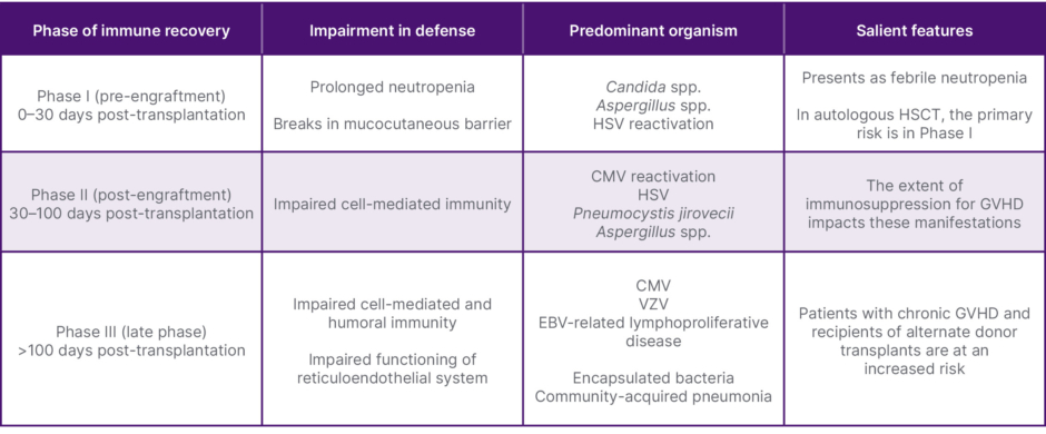
Table 1: Opportunistic infections in recipients of HSCT.18
CMV: cytomegalovirus; EBV: Epstein–Barr virus; GVHD: graft-versus-host disease; HSCT: hematopoietic stem cell transplantation; HSV: herpes simplex virus; VZV: varicella-zoster virus.
Compared to allogeneic stem cell transplantation, autologous HSCT has a lower risk of infectious complications; most infections in autologous HSCT occur during neutropenia or within the first few months after transplantation, but the manipulation of the graft might lead to an increased risk of infections.19 There have been attempts at reducing the risk of relapse in autologous HSCT with the use of cluster of differentiation (CD)34-selected peripheral blood stem cells, but the use of such grafts were associated with an increased risk of viral and bacterial infections.20 There was a statistically insignificant increase in the risk of fungal infection with the use of CD34-selected peripheral blood stem cells in an analysis by Crippa et al.21 The spectrum of individual pathogens associated with OI in patients undergoing HSCT will be discussed subsequently.
Opportunistic Bacteremia in HSCT
During the late transplant period, poor antibody and cell mediated immunity leads to a significant risk for infections with encapsulated bacteria, which can lead to development of pneumonia and/or meningitis. There is also an increasing occurrence of multidrug-resistant (MDR) species in the post-transplant setting. Prospective surveillance in a Brazilian study found bacteremia in 27% of patients, with 37% of those infections being attributed to MDR species. These MDR species can be acquired early through translocation of gut bacterial flora early during HSCT.22,23 Surveillance cultures have been used to determine the prevalence of MDR colonization in the endogenous flora of HSCT recipients: 50% of the nasal and axillary swabs of patients with Staphylococcus aureus isolates were methicillin resistant, as reported by Bhat et al.24 In surveillance fecal cultures, Korula et al.25 had seen a prevalence of drug-resistant colonization in 50% of the adult recipients of transplants and in >60% of the pediatric recipients.25 Prior colonization by carbapenem-resistant enterobacteriaceae (CRE) increases CRE-related blood stream infections, which are associated with high mortality.26 Consensus recommendations for the monitoring of CRE colonization as a part of the microbiological evaluation of potential HSCT recipients have been proposed in transplant centers with significant known CRE spread, in view of the poor prognosis associated with the infection.27
Reactivation of Mycobacterium tuberculosis has historically been reported to have low incidence in the post-transplant setting, but there have been reports of reactivation of latent pulmonary tuberculosis leading to severe infections in endemic areas. Non-tuberculous Mycobacteria (NTM) are ubiquitous environmental organisms, and the incidence of NTM in recipients of allogeneic HSCT (alloHSCT) has been found to be increasing, and in countries with lower incidence of M. tuberculosis, NTM infections predominate.28 The incidence of NTM among the recipients of alloHSCT has been found to range from 0.4–4.9%. Hirama et al.29 had looked at the characteristics of pulmonary disease after alloHSCT, and had found Mycobacterium avium to be the most common (50%) followed by Mycobacterium fortuitum (20%), Mycobacterium xenopi (15%), Mycobacterium abscessus (15%), and Mycobacterium gordonae (5%).23,28,29 Drug interactions between antibiotics and immunosuppressants complicate the management of NTM in recipients of alloHSCT, but it appears to be manageable in patients who are able to tolerate the antibiotic therapy, with up to 62% of patients experiencing resolution of symptoms.28,29
Opportunistic Viral Infections in HSCT
Viral infections after HSCT can be either secondary to reactivation of a latent infection or episodic events that are acquired by exposure. Cytomegalovirus (CMV), human herpesvirus 6, and varicella-zoster virus (VZV) represent viral infections that arise from reactivation, while respiratory syncytial virus, parainfluenza, influenza, and adenovirus, among others, are episodic in nature. Understanding this difference plays an important role in preventive strategies for these infections.
CMV infections have been a historically important cause of morbidity and mortality in HSCT recipients. It is common during the early pre-engraftment period, but with concomitant graft-versus-host disease (GVHD), it can be a significant cause of morbidity in the late transplant setting as well. CMV reactivation in seropositive recipients is the major cause of CMV-related infections, but primary infection of a seronegative recipient by transfusion of unfiltered blood or unmanipulated marrow from seropositive donors has been observed.30 The CMV seropositivity of the donor and/or recipient was found to have differing impacts on survival with respect to the type of donor. With partially T cell-depleted grafts in matched related donors, CMV seropositivity was not found to impact overall survival (OS) or treatment-related mortality (TRM), but in matched unrelated donors the patient seropositivity was found to impact both.30 The role of seropositivity in the donor has been less clearly defined. A European Society for Blood and Marrow Transplantation (EBMT) study had found no difference in OS and TRM in human leukocyte antigen-matched donors, but a significant impact on outcome in unrelated donors.31 A seropositive donor in an unrelated transplant was associated with improved event-free survival and reduced TRM. This phenomenon has been hypothesized to be explained by both a reduction in the rates of CMV reactivation, as well as the immune suppressive effects of CMV itself and associated concomitant infections.9,30,31
Herpes simplex virus (HSV) is an important pathogen in patients who develop neutropenia and mucositis. The reactivation rates without prophylaxis range from 60–80% in recipients of HSCT and patients with acute leukemia undergoing induction therapy.9 Disseminated infection is rare, but reactivation is frequently associated with increased pain and a decreased ability to maintain oral hygiene, leading to frequent superinfections.9
Impaired cell-mediated immunity is the principal risk factor for VZV reactivation. In the immunocompromised host, VZV reactivation can lead to more severe infections and a higher risk of complications, including central nervous system complications.32 The herpes zoster virus incidence has ranged from 37.2–56.1%, with an increased incidence noted with increasing age.9,32
Adenoviruses can lead to severe disseminated disease secondary to poor T cell reconstitution and GVHD, with the disease manifestations mimicking GVHD.23 The Polyoma virus family includes the BK virus, which is present as a latent infection in the urothelial cells of >90% of the population.23 It manifests as hemorrhagic cystitis. There is a lack of effective preventive strategies for both of these viruses.23
Prophylactic strategies have been established for several viral infections in immunocompromised patients. In case of CMV infections, the prophylactic strategy revolves around routine surveillance by PCR and initiation of pre-emptive therapy with ganciclovir or valganciclovir. The use of letermovir as a prophylactic strategy in recipients of allogeneic transplant who are CMV positive is also indicated.9 Prophylaxis for HSV is indicated based on the risk of these infections, and is indicated for the duration of immunosuppression; patients undergoing an allogeneic transplantation with severe GVHD should be considered for a longer duration of prophylaxis.9 Acyclovir, valacyclovir, and famciclovir are agents indicated for early prophylaxis.9 The same agents are indicated for prophylaxis against VZV infections, with prophylaxis being considered for 6–12 months post autologous transplantation and for at least 1 year after allogeneic transplantation.9
Opportunistic Fungal Infections in HSCT
Invasive fungal diseases (IFD) are an important cause of morbidity and mortality in patients undergoing HSCT, and the principal risk factor is prolonged and profound neutropenia, along with mucosal injury and the use of corticosteroids; the use of central venous catheters is a risk factor for candidemia.33 The incidence of IFD is higher than 10% in patients with acute myeloid leukemia, relapsed leukemia, and after allogeneic HSCT, while the risk is significantly lower (<5%) after autologous HSCT.33 The case fatality rates associated with IFD ranged from 20–70% in several case series.33 Yeast (most commonly Candida spp.) and mold infections are manifested by persistent or recurrent fever in patients with prolonged profound neutropenia. Candida spp. are colonizers of human mucosal surfaces, and infections can result from the breakdown of the mucosal barriers. Candida albicans is the most common species isolated from bloodstream infections, but there has been a trend toward an increase in the prevalence of non-albicans species, such as Candida krusei and Candida glabrata, which are intrinsically resistant to fluconazole.16,34 Candida parapsilosis is a non-Albicans Candida species that is known to adhere to the surface of catheters and lead to candidemia.16,33,34
Neutropenia remains the most important risk factor for invasive aspergillosis (IA), and there is a reported second period of risk that corresponds with the occurrence of GVHD. Other risk factors include the use of a T cell-depleted or CD34-selected stem cell products and the receipt of corticosteroids. The occurrence of CMV reactivation after Day 40 has been linked to an increased risk of IA; other respiratory infections (parainfluenza virus, respiratory syncytial virus) after Day 40 have also been associated with an increased risk of IA.35 The occurrence of IFD in chronic GVHD has been reported to be as high as 39%. Overall, outcomes with IA have been poor regardless of the time of onset of the infection, with a 30% survival at 3 months after diagnosis.33,35,36
Prophylactic antifungal therapy has helped to reduce fungal infection-related and all-cause mortality; the use of fluconazole has shown superiority over placebo in preventing fungal infections caused by Candida species, but has no role in the prevention of invasive molds.37 Fluconazole was compared with voriconazole in a randomized trial, which showed a trend toward a reduced incidence of IA with the use of voriconazole, without a statistically significant impact on OS.38 Posaconazole was also compared with fluconazole and was found superior in the prevention of IA.9,36
Pneumocystis jirovecii is a globally ubiquitous colonizing fungus that can colonize human pulmonary alveoli. Vulnerability to P. jirovecii pneumonia (PJP) is due to impaired T cell immunity in immunocompromised patients.Trimethoprim/sulfamethoxazole can be used as a preventive strategy, with prophylaxis continued throughout the period of immunosuppression.18,39
OI WITH NEWER THERAPIES
The armamentarium of therapeutic agents has advanced greatly over the past few decades, and with the advent of targeted therapies and cellular therapies, including CAR-T cell therapy and bispecific T cell engagers, along with the widespread use of checkpoint inhibitors in a variety of indications, there are newer infectious risks associated with these novel therapies.
OI with Checkpoint Inhibitors
ICIs that work by blocking key immune checkpoints, such as programmed cell death protein 1 (PD-1), programmed death-ligand 1 (PD-L1), and cytotoxic T-lymphocyte associated protein 4 (CTLA4), lead to a state of immune dysregulation rather than the classical immune suppression seen with cytotoxic chemotherapy. A consensus statement by the European Society of Clinical Microbiology and Infectious Diseases (ESCMID) study group had stated that CTLA4 and PD-1/PD-L1 targeted agents do not inherently raise the risk of infections, but the occurrence of immune-related adverse events could require additional immunosuppression that can predispose to OIs.40 At the level of prospective randomized trials with the use of these agents, the rate of reported infections has not been significant. Landmark trials have reported an incidence of neutropenia of Grade 3 or greater ranging from 2–16%.41,42 Morelli et al.43 proposed a framework for classification of infections associated with the use of ICI, dividing them into two categories: immune suppression and dysregulated immunity.44
Table 2 summarizes some of the reports of infectious complications associated with the use of ICIs. The overall incidence of infectious complications with the use of ICIs has been significantly lower when compared to cytotoxic chemotherapy. Fujita et al.,56 in a review of 167 patients with non-small cell lung cancer receiving nivolumab, documented infectious diseases in 19.2%, with 78% of them being bacterial. In a characterization of infectious complications following the use of ICIs, Kanjanapan et al.57 had seen 27% of the patients developing infections post-ICI therapy, which was non-significant compared to the pre-ICI rates. Both of these analyses did not attribute any added risk to the use of corticosteroids or immunosuppression.56,57 del Castillo et al.50 reported an infection rate of 7.3% in a large review of 740 patients, while also demonstrating an increased use of corticosteroids as a risk factor for the occurrence of infection.50 In a large retrospective analysis, Shah et al.53 had reported the incidence of OI to be 7% among patients receiving steroids for immune-related adverse events. The same analysis also found that the use of PJP prophylaxis was associated with a higher rate of breakthrough infection, thus questioning the role of prophylaxis in this patient population.53
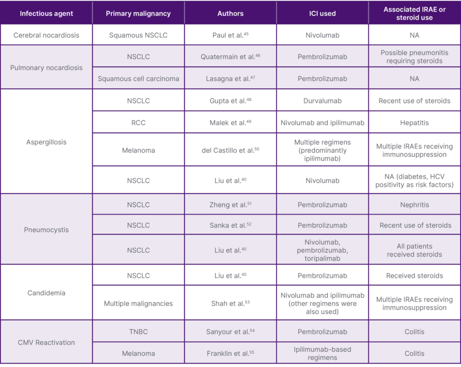
Table 2: Reports of OI in association with the use of ICIs.
CMV: cytomegalovirus; HCV: hepatitis C virus; ICI: immune checkpoint inhibitors; IRAE: immune-related adverse events; NA: not applicable; NSCLC: non-small cell lung cancer; OI: opportunistic infection; RCC: renal cell carcinoma; TNBC: triple negative breast cancer.
OI with Other Targeted Therapies
The reported infectious complications associated with other novel targeted therapies are summarized in Table 3.
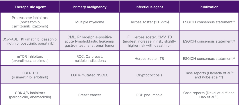
Table 3: Summary of reported evidence of infections with the use of novel targeted agents.
Ca: carcinoma; CDK 4/6: cyclin-dependent kinases 4 and 6; CML: chronic myeloid leukaemia; CMV: cytomegalovirus; EGFR: epidermal growth factor receptor; ESCMID: European Society of Clinical Microbiology and Infectious Diseases; ESGICH: ESCMID Study Group for Infections in Compromised Hosts; IFI: invasive fungal infections; PCP: pneumocystis pneumonia; RCC: renal cell carcinoma; TB: tuberculosis; TKI: tyrosine kinase inhibitor.
OI with CAR-T Cell Therapy and Bispecific Antibodies
CAR-T cell therapy has been a breakthrough in the management of several hematological malignancies, and there is a growing understanding of the complications associated with the use of this novel therapy.
Several factors influence the risk of infections in a patient undergoing CAR-T cell therapy, both in the pre-CAR-T infusion period and in the post-infusion period. Figure 1 summarizes the risk factors in both phases of treatment. Prolonged cytopenias after CAR-T cell therapy, now called immune effector cell-associated hematotoxicity, characterized by neutropenia persisting 30–90 days after CAR-T cell therapy, are a recognized risk factor for infections.63 Hypogammaglobulinemia has been reported in 62% of patients receiving anti-CD19 CAR-T cells and up to 75% of patients receiving anti-B cell maturation antigen CAR-T cells 1 year after the infusion.63,64
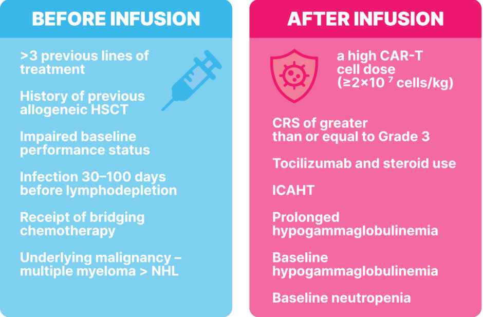
Figure 1: Risk factors for infections in recipients of CAR-T cell therapy.
CRS: cytokine release syndrome; HSCT: hematopoietic stem cell transplantation; ICAHT: immune effector cell-associated hematotoxicity; NHL: non-Hodgkin lymphoma; PS: performance status.
The spectrum of infections seen in the population of patients receiving CAR-T cells has been explored. Kambhampati et al.65 had found the rate of severe infections to be ~9%, while it was 3% in a study by Hill et al.11 The majority of the infections were viral (53%), followed by bacterial infections (40%), in the analysis by Kambhampati et al.,65 and Hill et al.11 had found bacterial infections to be more common in the early phase (first 28 days post-CAR-T cell therapy). In the same study, viral infections were found to be more common in the late phase (29–90 days post-CAR-T cell therapy).11,65 Both of these studies had found the incidence of mold infections to occur at significantly lower rates in this patient population.11,65
Bispecific antibodies (BsAb) bind to a target on the malignant cell, with simultaneous binding to an immune effector cell.
The use of BsAbs has led to improved survival in several hematological malignancies. Approximately one-third of the patients receiving these therapies can have prolonged cytopenias beyond 30 days of treatment. Hypogammaglobulinemia and lymphopenia following BsAb therapy are also recognized risk factors for infections. Other risk factors for infections, such as the inherently higher risk associated with the underlying condition and the management of cytokine release syndrome with steroids and tocilizumab, have also been implicated. In a pooled analysis of the use of BsAbs in multiple myeloma, Mazahreh et al.66 had shown a 4.2% incidence of PJP and a CMV reactivation rate of 8.0%. There have been consensus statements regarding anti-infectious prophylaxis in the setting of BsAbs, which can be summarized
as follows:64,66,67
- IVIg replacement is recommended in the following situations:
• Patients with Ig levels <400 mg/dL
• Patients who have experienced ≥2 severe recurrent infections by encapsulated bacteria regardless of the Ig level
• Patients with life-threatening infections
• Patients with documented bacterial infection with no or insufficient response to antibiotic therapy - Antiviral prophylaxis with acyclovir is recommended in all patients with relapsed refractory myeloma on treatment.
- General anti-bacterial prophylaxis is not recommended, and the treatment of microbial colonization is not recommended.
- Anti-PJP prophylaxis is recommended for all patients.
- Routine antifungal prophylaxis for other fungal infections is not recommended.
CONCLUSION
OIs are an important cause of mortality and morbidity in patients with cancer. There has been an evolution in the landscape of OIs seen, owing to both the changing treatment modalities and the use of prophylactic strategies. A continuing understanding of the evolution of pathogens and the recognition of risk factors associated with newer modalities of treatment will be vital to tailoring our diagnostic and therapeutic approaches in cases of suspected OI. Prophylactic strategies to mitigate the risk of OI in the setting of these newer therapies are evolving as we better understand the risks associated with them. At the same time, a matter of significant concern is the appearance of more resistant species of pathogens, which pose a significant challenge. Stewardship programs to mitigate indiscriminate use of antibiotics and other strategies to mitigate the risk of MDR species are the need of the hour.