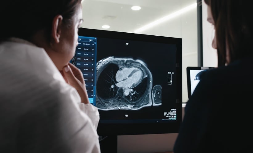
ARTIFICIAL intelligence-derived measurements of heart function showed a small improvement in mortality prediction compared with clinician measurements in a study of more than 27,000 patients.
The research, compared left-ventricular ejection fraction measured from cardiac MRI scans. The findings support further investigation of automated measurement with clinician oversight, although they do not demonstrate that using AI improves patient outcomes.
Comparing Measurements Across Two Countries
The retrospective, multicentre study included 27,382 patients who underwent cardiac MRI in the UK and USA. Most scans came from Barts Heart Centre, London, with an additional cohort from the University of Pittsburgh Medical Centre.
Researchers applied AI segmentation to the scans and compared the prognostic value of automated and clinician-derived measurements. During a median follow-up of 5.6 years, 3,458 patients died.
AI-derived ejection fraction provided better statistical model fit and slightly better discrimination between patients with different mortality outcomes. After adjustment for age and sex, the C index was 0.781 for AI measurements and 0.777 for clinician measurements, indicating a modest difference.
Faster Analysis With Expert Review
Ejection fraction is widely used to guide decisions about heart failure treatment, implanted devices and other interventions. Improving the consistency and efficiency of its measurement could therefore have practical relevance across imaging services.
According to the researchers’ institutional summary, automated analysis took approximately 2–3 seconds per patient. Previous work by the team found that expert clinician analysis typically took around 13 minutes. This comparison suggests a potential efficiency benefit, although it was not a direct measurement of time saved across an entire clinical service.
The researchers also emphasised the continuing need for human oversight. Automated errors were uncommon, but clinicians remained important for verifying measurements and interpreting findings within the wider clinical picture.
Establishing Clinical Value
The central result concerns prediction: AI measurements were more closely associated with subsequent mortality. That does not establish that adopting the tool changes treatment decisions appropriately or helps patients live longer.
Prospective evaluation would help determine whether the small statistical improvement translates into useful changes in care. It would also allow services to assess how automated measurements perform alongside clinician review in routine workflows.
For radiology teams, the findings provide a rationale for evaluating AI as an initial measurement tool. Its clinical value will depend on reliable quality assurance, appropriate review and evidence that faster analysis supports effective patient management.
Reference
Joy G et al. Performance of artificial intelligence versus clinician left-ventricular segmentation of cardiac MRI for mortality prediction: a retrospective, multicentre, prognostic study. Lancet Digit Health. Published online 2 November 2026. doi:10.1016/j.landig.2026.101042.